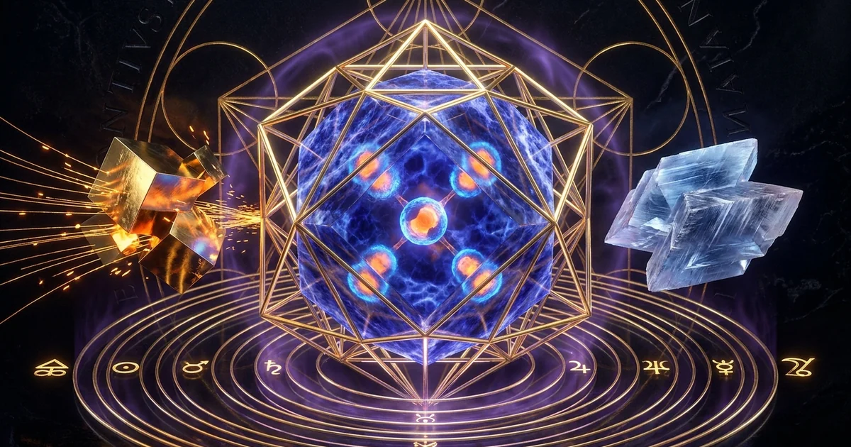
Lapis Lazuli Properties: Geology & Crystalline Resonance
Mineral Classification & Crystallographic Thesis: The Tripartite Metamorphic Assembly
Metamorphic Petrogenesis and Skarn Metasomatism
Lapis lazuli occupies an anomalous position in mineralogical taxonomy: it is not a distinct mineral species, but an unfoliated contact-metamorphic rock whose macroscopic properties arise from a petrogenetic paragenesis dominated by lazurite, calcite, and pyrite. The genesis of lapis lazuli is tied to high-grade metasomatic events, specifically pyrometasomatic contact skarns where magma of granitic-to-syenitic composition intrudes into sulfur-bearing dolomitic marbles and limestone formations. The archetype of this geological crucible is the historic Sar-e-Sang deposit situated along the Kokcha River Valley within the Badakhshan Province of northeastern Afghanistan. In these deep-seated crustal zones, amphibolite-to-granulite facies metamorphism operates under temperatures typically exceeding 600°C and pressures of several kilobars.
During these metasomatic interactions, silica- and alkali-rich volatile phases emanating from crystallizing felsic plutons permeate the surrounding carbonate rocks. This influx instigates decarbonation reactions, introducing sodium, silicon, and aluminum while mobilizing volatile sulfur compounds into the host matrix. Rather than producing standard calc-silicate skarn assemblies, such as grossular-andradite garnets or diopside, the intense activity of sodium and sulfur suppresses the stabilization of ordinary plagioclase feldspars. Instead, this thermodynamic environment drives the crystallization of feldspathoids. The resulting metamorphic rock (lazurite, pyrite, calcite) exhibits high structural heterogeneity, where the spatial convergence of these mineral phases creates complex chemical and electrical gradients at the grain boundaries. Understanding this petrogenetic origin is essential for analyzing the lapis lazuli crystal properties geology resonance profile, as the material’s macroscopic vibrational and dielectric properties are direct consequences of its multi-phase assembly.
Metasomatic Reaction Scheme:
Dolomitic Marble + Magmatic Volatiles (Si, Al, Na, S)
    ---> (Na,Ca)₈(AlSiO₄)₆(S,SO₄,Cl)₁₋₂ [Lazurite]
       + CaCO₃ [Calcite Recrystallization]
       + FeS₂ [Pyrite Nucleation]
       + Accessory Silicates (Diopside, Forsterite, Sodalite)
The localized structural order of the primary feldspathoid host lattice dictates the physical framework of the stone. This framework operates under spatial constraints modulated by adjacent accessory phases, whose mechanical boundaries generate localized stress fields throughout the bulk material.
The Sodalite-Group Host: Lazurite Framework Structure
The primary mineral phase defining the chromatic and structural character of lapis lazuli is lazurite, an aluminosilicate mineral belonging to the sodalite group of feldspathoids with the idealized chemical formula (Na,Ca)₈(AlSiO₄)₆(S,SO₄,Cl)₁₋₂. In the context of solid state crystallography, lazurite adopts a three-dimensional framework comprised of alternating silicon-dioxide-tetrahedra ($\text{SiO}_4$) and aluminum-oxygen tetrahedra ($\text{AlO}_4$) sharing all vertices, consistent with Löwenstein’s rule which forbids adjacent aluminum tetrahedra through $\text{Al-O-Al}$ linkages. This framework encloses a porous sub-nanometer topology known as the sodalite cage or $\beta$-cage, structurally configured as a truncated octahedron characterized by six four-membered rings and eight six-membered rings.
Lazurite crystallizes in the isometric (cubic) crystal system, characterized by the non-centrosymmetric space group $P\bar{4}3n$, though it frequently exhibits complex commensurate and incommensurate structural modulations caused by the spatial ordering of interstitial guest cations ($\text{Na}^+$, $\text{Ca}^{2+}$) and anions ($\text{S}_3^-$, $\text{SO}_4^{2-}$, $\text{Cl}^-$). As documented by Hogarth (1977) in his benchmark re-evaluation of the sodalite group, lazurite represents the sulfur-rich terminal member of this family, possessing a framework topology that accommodates polyatomic volatile species within its interstitial cavities. The structural integrity of the stone relies on the strength of its framework, where isotropic covalent-ionic bonds establish an average refractive-index typically ranging between 1.500 and 1.550, depending on the dynamic substitution of calcium for sodium and sulfate for sulfide within the aluminosilicate channels. For deeper inquiry into cage-framework solid mechanics, examine /crystals-materials/sodalite-lattice-dynamics.
- Crystal System: Isometric (Cubic)
- Space Group: $P\bar{4}3n$ (Space Group No. 218)
- Unit Cell Parameter: $a = 9.07 \text{ \AA} \text{ to } 9.09 \text{ \AA}$
- Unit Cell Volume: $V \approx 746.12 \text{ \AA}^3$
- Z (Formula Units per Cell): 1
- Mohs Hardness: 5.0–5.5 (pure lazurite phase); aggregate Mohs-hardness ranges from 5.0 to 6.5
- Aggregate Density: $2.70 \text{ g/cm}^3 \text{ to } 2.90 \text{ g/cm}^3$
- Framework Type: Sodalite ($\beta$-cage topology, truncated octahedron)
Source: Hogarth, D. D. (1977). ‘Classification and nomenclature of the sodalite group.’ The American Mineralogist, 62(3-4), 403-410.
Accessory Mineral Distribution: Pyrite Micro-Inclusions and Calcite Veining
Beyond its feldspathoid matrix, the lithological identity of lapis lazuli is established by its secondary and tertiary accessory phases: cubic iron disulfide (pyrite, $\text{FeS}_2$) and rhombohedral calcium carbonate (calcite, $\text{CaCO}_3$). Pyrite occurs as brassy, micro-crystalline to euhedral disseminated grains dispersed throughout the lazurite matrix. These inclusions nucleate during metasomatism as excess iron and sulfur segregate into localized isometric domains belonging to the space group $Pa\bar{3}$. Pyrite introduces localized zones of high mohs-hardness (6.0 to 6.5) alongside high electrical conductivity and distinct magnetic susceptibility profiles into the softer, insulating lazurite host matrix (hardness 5.0 to 5.5).
Spatial Microstructure of Lapis Lazuli:
+-------------------------------------------------------------+
|  [Lazurite Matrix: P4̄3n]       [Calcite Vein: R3̄c]          |
|  Insulating cage framework     Dielectric buffer zone       |
|  Mohs: 5.0-5.5                 Mohs: 3.0                    |
|             \                       /                       |
|              \                     /                        |
|        [Maxwell-Wagner Interfacial Boundary]                |
|                      /             \                        |
|                     /               \                       |
|  [Pyrite Micro-inclusion: Pa3̄]  [Radical Sulfur Cavity]    |
|  Semiconducting domain (0.95 eV) Unpaired spin (S=1/2)      |
|  Mohs: 6.0-6.5                 Paramagnetic center          |
+-------------------------------------------------------------+
Calcite, by contrast, manifests as white microcrystalline aggregates, interlaced bands, or structural micro-veining, crystallizing within the trigonal system (space group $R\bar{3}c$) with a lower Mohs hardness of 3.0. The spatial distribution of calcite dictates the mechanical friability and macro-porosity of the aggregate. These three distinct crystal structures form an intimate network of inter-phase boundaries. Because each mineral possesses contrasting dielectric, elastic, and galvanic potentials, their physical juxtaposition generates localized interfacial boundary-layer effects. These boundaries are fundamental to understanding the material’s macroscopic response to subtle electromagnetic and biofield fluctuations.
Lattice Geometry & Solid-State Physics: S₃⁻ Radical Chromophores and Dielectric Transduction
The Sodalite Cage: Sub-Lattice Trapping of Polysulfide Radical Anions
The visual presentation of lapis lazuli—its characteristic ultramarine coloration—originates from an unconventional, non-transition-metal chromophore system. The color is not produced by transition metal $d\text{-}d$ electronic transitions or charge transfers between octahedral cations. Instead, it arises from the quantum confinement of radical anions sequestered within the interstitial sodalite cages of the lazurite lattice. The principal chromophore responsible for the vivid 600 nm blue absorption is the trisulfur radical anion, $\text{S}_3^-$. The presence and fundamental dynamics of this chromophore were validated through resonance Raman spectroscopy and infrared analysis by Clark and Cobbold (1978), as well as Tarte and Preudhomme (1982).
“Resonance Raman spectra of ultramarine blue and natural lazurite demonstrate that the intense blue chromophore is definitively assigned to the fundamental symmetric stretching vibration ($\nu_1$) of the triatomic sulfur radical anion, $\text{S}3^-$, occurring at $548 \text{ cm}^{-1}$. The electronic absorption band centered near $600 \text{ nm}$ ($16,600 \text{ cm}^{-1}$) corresponds to the quantum-mechanically allowed $^2E’’ \leftarrow ,^2B_1$ transition within the $C{2v}$ molecular symmetry of the caged polysulfide species.” — Clark, R. J. H., & Cobbold, D. G. (1978). Inorganic Chemistry, 17(11), 3169-3174.
Within the rigid aluminosilicate framework, the triatomic $\text{S}_3^-$ radical anion is stabilized against oxidative degradation, thermal recombination, and atmospheric quenching. The molecular symmetry of the entrapped $\text{S}3^-$ radical conforms to a bent geometry ($C{2v}$ point group), possessing an open-shell electronic configuration with an unpaired electron situated in an antibonding $\pi^*$ molecular orbital. The electronic transition from the ground state $^2B_1$ to the excited state $^2E’'$ absorbs photon energies spanning yellow-to-red wavelengths ($\lambda \approx 580\text{–}620\text{ nm}$), leaving a transmission window in the deep blue and violet spectra. Trace quantities of the tetrasulfur radical anion ($\text{S}_4$) or the disulfur radical anion ($\text{S}_2^-$) can also occupy these cages, introducing subtle shifts toward reddish-purple or greenish-blue hues by modifying the localized band transitions.
Energy Level Diagram for Trapped S₃⁻ Radical:

                     [ ²E'' Excited State ]
                             ^
                             |
                             |  Electronic Absorption (λ ≈ 600 nm)
                             |  Deep-Red/Yellow Photons Absorbed
                             |
                     [ ²B₁ Ground State ]
                  (Unpaired electron, S = 1/2)
The aluminosilicate framework functions as an electrostatic trap. Cations such as $\text{Na}^+$ and $\text{Ca}^{2+}$ counterbalance the net negative charge of the sulfur radicals, while physical cage dimensions (free interior diameter $\sim 6.6\text{ \AA}$) inhibit translation. As a result, the radicals exist as an isolated array of quantum spins dispersed throughout an insulating solid-state matrix.
Dielectric Spectroscopy, Permittivity, and Conductivity Profiles
The multi-phase heterogeneity of lapis lazuli produces complex dielectric properties that contrast sharply with single-crystal silicates. When exposed to alternating electric fields, lapis lazuli exhibits significant frequency dispersion across its real ($\varepsilon’$) and imaginary ($\varepsilon’'$) dielectric-constant components. This dispersion is particularly pronounced within the low-frequency to radio-frequency spectrum ($10^2 \text{ Hz}$ to $10^7 \text{ Hz}$). This dielectric behavior is largely governed by the contrast between its constituent mineral phases: insulating lazurite, semi-conducting pyrite, and dielectric calcite.
Pyrite ($\text{FeS}_2$) possesses an indirect narrow bandgap of approximately $0.95 \text{ eV}$, functioning as an intrinsic semiconductor with elevated carrier concentrations and high electrical conductivity relative to its silicate surroundings. Its properties are detailed in /crystals-materials/pyrite-semiconducting-resonance. The lazurite framework, conversely, has a bandgap exceeding $4.5 \text{ eV}$ and behaves as an electrical insulator, though interstitial cations retain restricted hopping conductivity at elevated temperatures.
Calcite acts as a dielectric buffer characterized by low loss factors and an alternating-current permittivity of $\varepsilon_r \approx 8.0\text{–}8.5$. When an external electromagnetic field intersects these phase junctions, charge carriers migrate through the semiconducting pyrite domains but cannot cross the insulating lazurite-calcite interfaces. This leads to charge accumulation at the structural boundaries.
Lazurite: Framework Insulator & Chromophore
- Chemical Formula: $(\text{Na},\text{Ca})_8(\text{AlSiO}_4)_6(\text{S},\text{SO}4,\text{Cl}){1-2}$
- Symmetry: Isometric, $P\bar{4}3n$
- Electronic Bandgap: $\approx 4.5\text{–}5.0 \text{ eV}$ (Dielectric Insulator)
- Relative Permittivity ($\varepsilon_r$): $\approx 6.5\text{–}7.8 \text{ at } 100 \text{ kHz}$
- Dominant Mechanism: Polysulfide radical quantum transitions ($\text{S}_3^-$ at $600 \text{ nm}$); paramagnetic resonance; cation-hopping polarization along sodalite channels.
Pyrite: Semiconducting Inclusions
- Chemical Formula: $\text{FeS}_2$
- Symmetry: Isometric, $Pa\bar{3}$
- Electronic Bandgap: $\approx 0.95 \text{ eV}$ (Narrow-gap Semiconductor)
- Relative Permittivity ($\varepsilon_r$): Apparent $\varepsilon_r > 30\text{–}100$ (Low frequency, metallic-like screening)
- Dominant Mechanism: High mobile charge-carrier density; Maxwell-Wagner charge buildup; RF micro-diode rectification at silicate boundaries.
Calcite: Dielectric Matrix Buffer
- Chemical Formula: $\text{CaCO}_3$
- Symmetry: Trigonal, $R\bar{3}c$
- Electronic Bandgap: $\approx 6.0 \text{ eV}$ (Wide-gap Insulator)
- Relative Permittivity ($\varepsilon_r$): $\approx 8.0\text{–}8.5 \text{ at } 100 \text{ kHz}$
- Dominant Mechanism: Electric field damping; spatial impedance buffer; ionic polarization via localized rhombohedral lattice distortion.
This multi-component assembly is a classic physical model of Maxwell-Wagner-Sillars (MWS) polarization. As detailed in the study of /physics-electromagnetism/maxwell-wagner-polarization, charge accumulation at the interfaces creates large artificial dipole moments, leading to apparent low-frequency relative permittivity values ($\varepsilon_r$) that exceed $10^2$ to $10^3$ below $1 \text{ kHz}$. As the field frequency increases, these trapped carriers can no longer track the oscillating vector, leading to an abrupt decline in permittivity alongside a dielectric dissipation peak ($\tan \delta$) in the high kilohertz range.
Interfacial Polarizability and Acoustic Phonon Propagation
The complex assembly of lapis lazuli also alters the behavior of acoustic phonons—the quantized lattice vibrations traversing its interior. Because each component mineral displays different densities ($\rho_{\text{calcite}} \approx 2.71 \text{ g/cm}^3$, $\rho_{\text{lazurite}} \approx 2.4\text{–}2.5 \text{ g/cm}^3$, $\rho_{\text{pyrite}} \approx 5.01 \text{ g/cm}^3$) and distinct elastic stiffness tensors ($C_{ijkl}$), the propagation of acoustic wave packets through the rock is characterized by continuous impedance mismatching at the phase interfaces.
Acoustic Phonon Dispersion & Scattering at Mineral Boundaries:
-------------------------------------------------------------------------
Incident Phonon Wave Vector (k) --->
       |
       |===> Boundary [Lazurite / Pyrite]
       |     Impedance Mismatch (Z₁ != Z₂)
       |
       +---> Reflected High-Frequency Transverse Modes (Acoustic Phonons)
       +---> Interfacial Shear Strain Generation (Local Stress Field)
       +---> Transduced Evanescent Piezoelectric Potentials
-------------------------------------------------------------------------
High-frequency phonons traversing the stone experience Rayleigh and Mie scattering when encountering micro-scale pyrite inclusions and calcitic boundary planes. This structural scattering attenuates coherent transverse acoustic modes while converting primary acoustic waves into localized interfacial shear strain. This mechanical-to-dielectric coupling creates micro-scale spatial variations in electric fields through strain-induced interfacial polarizability. This behavior establishes lapis lazuli as an inhomogeneous, solid-state electro-acoustic transducer operating across both macroscopic mechanical domains and atomic-scale lattice networks.
Subtle Energetic Dynamics & Resonance Mechanics: Biofield-Lattice Transduction
Piezoelectric and Electrostrictive Contributions of Multi-Phase Interfaces
Assessing the electromechanical transduction properties of lapis lazuli requires isolating the individual crystal symmetries of its constituent phases. Ideal cubic lazurite crystallizes in the space group $P\bar{4}3n$, which lacks a center of inversion. Centrosymmetry is the definitive crystallographic factor that determines whether a mineral can support linear piezoelectricity. Consequently, the ideal single-crystal lazurite lattice permits a single non-zero piezoelectric tensor component: the shear coefficient $d_{14}$. In macroscopic specimens, however, lazurite rarely exhibits clean single-crystal orientations; it occurs instead as an aggregate of micro-twinned domains.
While pure cubic pyrite ($Pa\bar{3}$) and rhombohedral calcite ($R\bar{3}c$) possess inversion centers that preclude intrinsic, bulk linear piezoelectricity, the heterogeneous multi-phase system as a whole exhibits non-centrosymmetric boundary behavior. The primary electromechanical mechanism in lapis lazuli arises along its internal interfaces:
Interfacial Strain Generation Mechanism:
[ Bulk Elastic Stress ] ---> [ Modulus Mismatch: Lazurite/Pyrite/Calcite ]
                        ---> [ Localized Interfacial Shear (d₁₄ Activation) ]
                        ---> [ Electrostrictive Dipole Polarization ]
                        ---> [ Local Electric Field Generation ]
When mechanical or acoustic pressure waves deform the rock, the elastic modulus mismatch between pyrite ($K \approx 140\text{–}160 \text{ GPa}$) and the softer lazurite-calcite matrix ($K \approx 60\text{–}80 \text{ GPa}$) focuses shear stress directly along the structural grain boundaries. These boundaries contain uncompensated surface charges, broken coordinate bonds, and oriented radical centers. The combination of localized shear along lazurite’s $d_{14}$ tensor and non-linear electrostrictive effects within the calcite-pyrite boundary zones allows the stone to transduce ambient acoustic and mechanical fluctuations into localized electric potential gradients.
Biofield Electromagnetic Coupling: Upper-Dantian and Throat-Chakra Energetics
Within subtle field mechanics, lapis lazuli has long been aligned with the human communicative and cognitive subtle-energy-vortices: the Vishuddha (throat) and Ajna (third eye or upper-dantian) centers. Analyzed from a solid-state perspective, this traditional correlation maps to the dielectric properties of the rock and its capacity to interact with weak endogenous bio-electromagnetic emissions. The human nervous system, cardiovascular tree, and brain generate endogenous electromagnetic fields characterized by ultralow frequencies (ELF) ranging from $0.5 \text{ Hz}$ to several hundred Hertz, accompanied by high-frequency biophoton emissions centered in the visible and near-ultraviolet spectra.
Lapis lazuli interacts with these biological fields through a combination of its Maxwell-Wagner interfacial capacitance and the semiconducting behavior of its micro-pyrite inclusions. Dispersed throughout an insulating matrix, these pyrite inclusions function as distributed Schottky barrier junctions and point-contact diodes. This network can rectify high-frequency carrier waves, transducing ambient radio-frequency noise and bioelectromagnetic fluctuations into weak direct-current polarization fields.
Subtle Biofield Transduction Cascade:
+-------------------------------------------------------+
|  Endogenous Biofield / Neural Field Emission          |
+-------------------------------------------------------+
                           |
                           v
+-------------------------------------------------------+
|  Maxwell-Wagner Interfacial Charge Polarization       |
|  (Phase boundary accumulation at 10²–10⁵ Hz)          |
+-------------------------------------------------------+
                           |
                           v
+-------------------------------------------------------+
|  S₃⁻ Radical Spin Coherence / Paramagnetic Response   |
|  (Zeeman splitting; paramagnetic dipolar coupling)    |
+-------------------------------------------------------+
                           |
                           v
+-------------------------------------------------------+
|  Micro-Pyrite Rectification                           |
|  (Localized micro-junctions convert RF/ambient noise) |
+-------------------------------------------------------+
                           |
                           v
+-------------------------------------------------------+
|  Modulated Subtle Resonant Feedback to Biofield       |
+-------------------------------------------------------+This multi-phase transduction matches the subtle energetic dynamics associated with the Vishuddha chakra (focused on frequency articulation, vocal resonance, and linguistic coherence) and the Ajna center (associated with visual pattern processing and phase coherence). The stone’s structural components function as an impedance-matching network that bridges somatic electromagnetic currents with subtle biofield matrices.
Quantum Spin States in Sulfur Radicals as Subtle Coherence Catalysts
Beyond its bulk electrical interactions, the trisulfur radical anion ($\text{S}_3^-$) introduces quantum spin dynamics to lapis lazuli. Possessing an odd electron count, the trapped $\text{S}_3^-$ radical acts as a stable spin-1/2 ($S = 1/2$) paramagnetic center. In conventional diamagnetic aluminosilicates, all electron spins are paired within localized covalent bonds. The lazurite lattice, however, features an array of unpaired paramagnetic spins isolated inside the structural $\beta$-cages.
Under ambient magnetic conditions, these isolated sulfur spins experience Zeeman splitting, separating the energy levels of their spin-up and spin-down states. The long relaxation times of these localized radicals make them sensitive to weak external magnetic fluctuations:
Zeeman Splitting in the Trapped S₃⁻ Radical:

                      E + ½ g μ_B B  (m_s = +½, Spin Anti-parallel)
                    /
  Ground State ----
  (S = ½, B = 0)    \
                      E - ½ g μ_B B  (m_s = -½, Spin Parallel)

  Applied Field (B) --->
Because the $\text{S}_3^-$ radicals are physically shielded by the aluminosilicate cage, their quantum spin states exhibit long decoherence lifetimes relative to free radical species in solution. When subtle biofield vectors intersect this paramagnetic lattice, the spin centers undergo weak dipolar coupling. This spin interaction allows the stone to function as a solid-state coherence catalyst, aligning external electromagnetic micro-distortions with the stable spatial symmetry of the sodalite matrix. The interaction of these cubic frameworks with metaphysical archetypes is examined further in /sacred-geometry/metatrons-cube-mineral-lattices.
Historical Lapidary Lore & Traditional Lineage: From Inanna’s Descent to Renaissance Ultramarine
The Ancient Near East and Badakhshan Trade Corridors
The historical lineage of lapis lazuli is tied to the trade corridors linking the Hindu Kush mountains to the early urban centers of the Ancient Near East. Dating back to the fourth millennium BCE, the Sar-e-Sang mines in Badakhshan operated as the primary global source of this material. The stone was transported across the Iranian plateau to Mesopotamia, the Levant, and Egypt. In the visual and literary culture of Sumer, Akkad, and Babylonia, lapis lazuli (Akkadian: uqnû; Sumerian: za-gìn) was viewed not merely as a decorative gem, but as a condensed mineral manifestation of divinity, celestial authority, and sacred power.
Ancient Near Eastern Trade and Mystical Trajectory:
[ Badakhshan / Sar-e-Sang ] (Geological Metasomatic Origin)
            |
            v  (Overland Trans-Iranian Caravan Routes)
[ Mesopotamia: Ur, Uruk, Nippur ]
  - Inanna's Talismanic Adornment (Pectoral, Measuring Rod)
  - Royal Headdresses, Amuletic Seals, Cuneiform Reification
            |
            v  (Maritime & Desert Trade Arteries)
[ Dynastic Egypt ]
  - Flesh of the Gods (Ra's Hair, Khepri's Carved Carapace)
  - Eye of Horus Inlays, Pectoral Gold-Lapis Soldering
In the Sumerian myth of the Descent of Inanna (circa 2000 BCE), the goddess equips herself with lapis lazuli talismans before her journey into the underworld (Kur). She carries the lapis measuring rod and cord (E-EŠ₂) and wears lapis beads around her neck as talismans of authority. To the Sumerian priesthood, the stone carried ontological weight: its deep blue hue mirrored the night sky, while the dispersed gold pyrite flecks were seen as manifestations of the primary creative principle.
In Dynastic Egypt, lapis lazuli (khesbedj) was similarly regarded as the physical embodiment of divine flesh, specifically associated with the hair of the sun-god Ra. It was frequently carved into heart scarabs, Eyes of Horus (wadjet), and protective amulets embedded within funerary masks, including the pectoral arrays of Tutankhamun. The Egyptian temple priesthood utilized the stone to link physical rituals with subtle celestial energies, leveraging its structural stability to anchor protective spiritual vectors.
Classical Greco-Roman Lapidaries: Sappheiros of Theophrastus and Pliny
In Classical Greco-Roman lapidary literature, the nomenclature used for blue minerals diverged from modern mineralogical terminology. Authors of antiquity did not use the term “lapis lazuli” (a medieval Latin formulation adapted from the Arabic lāzaward and Persian lāžward). Instead, they designated this multi-phase rock as sappheiros ($\sigma\alphá\pi\varphi\varepsilon\iota\rho\text{o}\varsigma$). The modern sapphire (corundum, $\text{Al}_2\text{O}_3$) was largely unfamiliar to Mediterranean naturalists or was classified under different lapidary designations.
Theophrastus, writing in his fourth-century BCE treatise De Lapidibus (On Stones), provided one of the earliest surviving mineralogical descriptions of this material:
“The sappheiros is dark in color and not very different from the smaragdus; it possesses markings that appear golden, speckled throughout its surface, yet it is by nature an aggregate stone, formed of distinct parts that vary under the graver’s tool.” — Theophrastus. (ca. 315 BCE). De Lapidibus, Section 23. Translation by E. R. Caley & J. F. C. Richards, Ohio State University Press (1956).
Pliny the Elder elaborated on these observations in his Naturalis Historia (ca. 77 CE), providing structural details that distinguished the stone from other colored lapidary minerals:
“Sapphiros enim et aureis punctis collucet. Caeruleae et sapphiri, raroque cum purpura, optimaeque apud Medos; nusquam tamen perlucidae. Praeterea inutiles scalpturae interveniente nexu calcis.”
(Sappheiros also shines with golden spots. These stones are blue, rarely tinged with purple, and the finest specimens come from the country of the Medes; yet they are never transparent. Furthermore, they are unsuitable for engraving when interrupted by veins of calcite.) — Pliny the Elder. (ca. 77 CE). Naturalis Historia, Book XXXVII: Lapidary Materials and Gemstones (Trans. H. Rackham, Loeb Classical Library).
Pliny’s evaluation captures the multi-phase composition of lapis lazuli without modern analytical instrumentation. His observation that the stone is “refulgent with golden spots” (aureis punctis collucet) accurately identifies the dispersed cubic pyrite inclusions. Similarly, his caution that structural veins of calcite (nexu calcis) weaken its structural integrity highlights the mechanical challenges posed by inter-phase boundaries during carving and lapidary preparation.
Alchemical Fraughtness: The Extraction of Fra Angelico’s Ultramarine
The transition of lapis lazuli from an ornamental carving stone to an artistic medium occurred during the late medieval and early Renaissance periods through the extraction of ultramarine (azzurro oltremarino—“blue from beyond the sea”). Raw, powdered lapis lazuli yields an unstable, greyish-blue pigment if crushed directly, as the color intensity of lazurite is diminished by intergrown calcite and pyrite impurities. The process of isolating the lazurite framework required a multi-stage physical-chemical extraction method known as pastiglia levigation, documented in detail by Cennino Cennini in his fourteenth-century treatise Il Libro dell’Arte.
The Renaissance Pastiglia Levigation Method:
+-------------------------------------------------------+
| Pulverize Raw Lapis Lazuli Rock                       |
| (Lazurite + Pyrite + Calcite Matrix)                  |
+-------------------------------------------------------+
                           |
                           v
+-------------------------------------------------------+
| Knead into Molten Mastic Paste                        |
| (Pine Rosin, Beeswax, Gum Mastic, Linseed Oil)        |
+-------------------------------------------------------+
                           |
                           v
+-------------------------------------------------------+
| Submerge & Shear in Weak Alkaline Lye (K₂CO₃)         |
+-------------------------------------------------------+
        /                                       \
       /                                         \
      v                                           v
[ Hydrophilic Settling ]               [ Oleophilic Retention ]
Lazurite Cages Separate into Water     Calcite & Pyrite Cling to Paste
Yields Pure Ultramarine                Yields Ash Residual (*Cendres*)
This extraction method relied on differences in surface interfacial tensions among the constituent minerals. Finely pulverized lapis lazuli was kneaded into a warm paste of beeswax, pine resin, mastic, and linseed oil. After cooling, the paste was kneaded submerged in a weak aqueous solution of potassium carbonate (lye). The hydrophilic surfaces of the aluminosilicate lazurite cages allowed them to wet and precipitate into the water, while the calcite and pyrite clung to the oleophilic wax-resin mass.
The resulting pigment, used by Renaissance masters such as Giotto and Fra Angelico, retained the full quantum absorption of the $\text{S}_3^-$ radical anion. This material purity contributed to its high value, making ultramarine more expensive than gold across Renaissance Europe.
Practical Applications, Calibration & Safety Protocols: Handling, Grids, and Material Fragility
Geometric Alignment and Biofield Attunement Protocols
When deploying lapis lazuli within subtle energy grids, spatial balancing designs, or biofield calibration setups, practitioners must account for its heterogeneous crystallographic structure. Because natural lapis lazuli contains both paramagnetic centers ($\text{S}_3^-$) and semiconducting inclusions (pyrite), it responds anisotropically to directional energetic vectors. Alignment protocols must consider both the physical morphology of the specimen and its accessory mineral distribution.
Spatial Grid Alignment Matrix:
                    [ True North (Magnetic Field B₀) ]
                                   ^
                                   |
              +--------------------+--------------------+
              |                                         |
     [ Pyrite-Dense Facets ]                   [ Calcite-Rich Zones ]
     Aligned along North-South Vector          Oriented toward Grid Periphery
     Leverages localized magnetic permeability Provides spatial dielectric buffer
     and semiconducting RF rectification       Prevents field collapse across node
              |                                         |
              +--------------------+--------------------+
                                   |
                                   v
                    [ True South (Biofield Axis) ]
Optimal placement aligns specimens along north-south axes using facets with dense, visible pyrite inclusions. This orientation aligns the weak magnetic permeability of the iron disulfide phases with the Earth’s geomagnetic field vector, establishing a stable physical foundation for the surrounding field.
Calcite-rich sections are best positioned outward toward the periphery of the grid. This arrangement utilizes calcite’s lower dielectric constant as a natural buffer, shielding the high-permittivity lazurite core and preventing field dissipation across adjacent grid elements. When combined in sacred geometry grids, such as the dodecahedral and vector-equilibrium frameworks of Metatron’s Cube, lapis lazuli functions as a grounding node that anchors high-frequency acoustic and mental intentions into the physical environment.
Material Degradation: Chemical Vulnerabilities to Acids and Moisture
The mineralogical diversity that gives lapis lazuli its unique properties also introduces chemical vulnerabilities. The aggregate is susceptible to structural breakdown when exposed to common atmospheric contaminants, acidic solutions, and unsuitable cleaning methods. The structural weak point in the stone is the rhombohedral calcite phase ($\text{CaCO}_3$). Calcite reacts readily with weak acids—including dilute acetic acid (vinegar), citric acid, and sweat—undergoing rapid dissolution via carbon dioxide effervescence:
$$\text{CaCO}_3 + 2\text{H}^+ \longrightarrow \text{Ca}^{2+} + \text{H}_2\text{O} + \text{CO}_2 \uparrow$$
This chemical reaction produces surface pitting, strips the material of its polish, and weakens the underlying aluminosilicate framework.
Chemical Attack Vulnerabilities:
+-------------------+-----------------------------------+------------------------------------+
| Phase             | Chemical Trigger                  | Degradation Outcome                |
+-------------------+-----------------------------------+------------------------------------+
| Calcite (CaCO₃)   | Acidic exposure (pH < 6.0)        | Immediate dissolution, pitting     |
| Pyrite (FeS₂)     | Moisture + Oxygen (H₂O + O₂)      | Sulfuric acid release, rust        |
| Lazurite Matrix   | Ultrasonic acoustic cavitation    | Micro-fracturing along twinning    |
+-------------------+-----------------------------------+------------------------------------+
Concurrently, the micro-inclusions of iron disulfide (pyrite, $\text{FeS}_2$) are prone to chemical oxidation when exposed to moisture and oxygen:
$$2\text{FeS}_2 + 7\text{O}_2 + 2\text{H}_2\text{O} \longrightarrow 2\text{Fe}^{2+} + 4\text{SO}_4^{2-} + 4\text{H}^+$$
This oxidation reaction releases trace amounts of sulfuric acid directly into the surrounding stone matrix. The resulting internal acidity accelerates the decomposition of nearby calcite and breaks down the sodalite cages, stripping the stone of its blue hue and leaving dull brown stains of iron hydroxide (limonite/rust).
Consequently, lapis lazuli must never be cleaned with steam or subjected to ultrasonic cleaning. The high-frequency shockwaves generated by ultrasonic cavitation strip away micro-calcite veining and induce micro-fracturing along lazurite’s structural twinning planes.
Toxicity Risks in Elixir Production and Leaching Dynamics
The chemical instability of lapis lazuli poses severe toxicological risks if the stone is introduced directly into water intended for consumption. Producing direct gem elixirs by soaking raw or polished lapis lazuli in water introduces hazards stemming from both the sulfur-bearing aluminosilicate framework and the sulfide accessory phases.
- Direct Gem Elixir Prohibition: Lapis lazuli must NEVER be immersed directly in drinking water, consumable solutions, or culinary matrices.
- Hydrogen Sulfide Release: Exposure to low-pH gastric acids or slightly acidic water breaks open the unstable polysulfide sodalite cages, hydrolyzing the sulfur content and releasing toxic, volatile hydrogen sulfide gas ($\text{H}_2\text{S}$): $$\text{S}_3^{2-} + 2\text{H}^+ \longrightarrow \text{H}_2\text{S} \uparrow + , 2\text{S}^0$$
- Heavy Metal Leaching: Accessory pyrite inclusions frequently contain substitutional impurities of arsenic, nickel, and cobalt within their lattices. During the oxidation of pyrite in aqueous solutions, these toxic metals can leach directly into the fluid.
- Safe Preparation Method: Energetic elixirs must be prepared exclusively through the Indirect Method, placing the specimen inside a sealed, sterile glass container before immersion in an outer water vessel.
- Cleaning Protocols: Clean the stone using dry methods only: employ soft microfiber cloths, brief acoustic cleansing (tuning forks), or dry placement on clean quartz substrates. Prohibit all salt, water, steam, or chemical detergents. Consult /crystals-materials/toxic-minerals-elixir-safety for comprehensive toxicological protocols.
These leaching pathways can be activated even by brief exposure to pure water, as structural micro-pores retain moisture long after immersion. Over time, trapped water facilitates internal galvanic reactions between the pyrite and lazurite domains, destabilizing the stone from within.
Synthesis & Advanced Diagnostics: Spectroscopy, Authenticity, and Energy Dynamics
Spectroscopic Differentiation: Distinguishing Lazurite from Sodalite, Hauyne, and Shattuckite
Given the market prevalence of dyed substitutes and related mineral phases, definitive mineralogical identification requires non-destructive spectroscopic analysis. Standard lapidary testing methods, such as assessing the aggregate refractive index via the spot method ($\sim 1.50\text{–}1.52$) or measuring specific gravity ($2.70\text{–}2.90 \text{ g/cm}^3$), provide general classification data but often fail to separate lazurite from other sodalite-group minerals or dyed carrier rocks like magnesite.
Comparative Raman Spectral Signatures:
1. Lapis Lazuli (Lazurite Phase):
   [548 cm⁻¹: S₃⁻ ν₁ Fundamental] <--- Strong, Sharp
   [1096 cm⁻¹: S₃⁻ 2ν₁ Overtone]  <--- Distinct Diagnostic Peak
   [1640 cm⁻¹: S₃⁻ 3ν₁ Harmonic]  <--- Trace Component

2. Sodalite (Chlorine Dominant):
   [256 cm⁻¹: Lattice Ring Mode]
   [460 cm⁻¹: Si-O-Si Band]
   (Absence of 548 cm⁻¹ S₃⁻ resonance)

3. Dyed Magnesite / Howlite:
   [1094 cm⁻¹: CO₃²⁻ Carbonate Stretch]
   (Broad organic dye bands at 1200–1600 cm⁻¹)
Raman spectroscopy remains the definitive laboratory standard for identifying genuine lapis lazuli. The presence of the $\text{S}_3^-$ radical anion within the sodalite cage yields a dominant Raman shift at $548 \text{ cm}^{-1}$, corresponding to the fundamental symmetric stretching vibration ($\nu_1$) of the triatomic sulfur molecule. This peak is accompanied by an overtone series at $1096 \text{ cm}^{-1}$ ($2\nu_1$) and $1640 \text{ cm}^{-1}$ ($3\nu_1$).
Sodalite ($\text{Na}_8(\text{AlSiO}_4)_6\text{Cl}_2$) lacks this $548 \text{ cm}^{-1}$ signature because it contains chlorine rather than polysulfide radical groups. Hauyne exhibits an overlapping peak but typically features an intense band at $990 \text{ cm}^{-1}$ corresponding to sulfate ($\text{SO}_4^{2-}$) framework groups. Secondary blue copper silicates like shattuckite ($\text{Cu}_5(\text{SiO}_3)_4(\text{OH})_2$) are differentiated by copper-hydroxyl vibrational bands located beyond $3000 \text{ cm}^{-1}$.
Synthetic Ultramarine and Reconstituted Imitations: Raman Fingerprinting
The widespread production of synthetic ultramarine (invented independently by Guimet and Gmelin in 1828) alongside modern polymer-bonded “reconstituted lapis” presents challenges for authenticity testing. Synthetic ultramarine contains the same $\text{S}_3^-$ radical chromophore as natural lazurite, meaning it produces identical Raman resonance shifts at $548 \text{ cm}^{-1}$. Differentiating natural lapis lazuli from synthetic or composite imitations requires looking beyond the primary chromophore to evaluate the rock’s overall mineral matrix.
Natural lapis lazuli always retains traces of its contact skarn petrogenesis. Petrographic thin-section microscopy and micro-Raman mapping reveal natural accessory silicates, including diopside, forsterite, and wollastonite, alongside irregular, naturally nucleated calcite and pyrite grains. Synthetic ultramarine, when bound in modern acrylic or epoxy matrices, displays a uniform groundmass without mineralogical zoning. Furthermore, polymer-bonded imitations show distinct organic Raman signatures from polymer binders (such as $\text{C-H}$ stretching bands between $2800$ and $3100 \text{ cm}^{-1}$) and lack natural Maxwell-Wagner interfacial polarization, as the insulating synthetic resins isolate the individual mineral grains.
Authenticity Assessment Profile:
+------------------------------------+---------------------------------------+
| Natural Lapis Lazuli               | Reconstituted / Synthetic Imitation   |
+------------------------------------+---------------------------------------+
| Multiphase mineral matrix          | Uniform polymer or epoxy matrix       |
| Accessory diopside & wollastonite  | Complete lack of accessory skarn phases |
| Heterogeneous Maxwell-Wagner effect| Weak, non-dispersive dielectric curve |
| Sharp 548 cm⁻¹ + 1096 cm⁻¹ peaks   | Chromophore present, but resin bands  |
| Natural irregular pyrite graining  | Metallic dust, brass, or absent pyrite|
+------------------------------------+---------------------------------------+
Vibrational Longevity and Subtle Coherence Retention
Maintaining the long-term resonance properties of lapis lazuli requires protecting its delicate lattice architecture from environmental stress. The stone’s subtle field dynamics rely on the charge stability of its interstitial sulfur radicals, the mechanical integrity of its phase boundaries, and the preservation of its water-free aluminosilicate channels.
To clear accumulated environmental charges and maintain the dielectric integrity of the lapis lazuli matrix:
- Acoustic Restabilization: Expose the stone to coherent acoustic vibrations using quartz singing bowls or aluminum tuning forks calibrated to $528 \text{ Hz}$ or $432 \text{ Hz}$. Acoustic vibrations relieve localized mechanical stresses along the lazurite-pyrite grain boundaries through structural relaxation.
- Geometric Substrate Coupling: Rest the stone on a dry bed of untreated natural selenite (gypsum) or optical quartz for four to six hours. This provides a clean dielectric substrate that dissipates accumulated static surface charges without introducing moisture.
- Thermal Environment Control: Avoid exposing the stone to direct sunlight or temperatures above $60^\circ\text{C}$. Excessive heat causes thermal expansion mismatch between pyrite and calcite, producing irreversible micro-fracturing along their mutual boundaries.
Subjecting the material to thermal shock causes contrasting expansion rates between the pyrite inclusions ($\alpha \approx 8.4 \times 10^{-6} \text{ K}^{-1}$) and the lazurite matrix ($\alpha \approx 6.5 \times 10^{-6} \text{ K}^{-1}$). This thermal disparity produces localized boundary detachment, disrupting internal Maxwell-Wagner interfacial charge mobility and degrading the stone’s capacity to transduce subtle biofield frequencies.
Frequently Asked Questions: Mineralogical Precision and Energetic Handling
Lapis Lazuli Mineral Composition and Hardness Nuances
Why is lapis lazuli classified as a rock rather than a distinct mineral species?
A mineral is defined crystallographically as a naturally occurring, homogeneous, inorganic solid with a definite chemical composition and an ordered internal atomic arrangement. Lapis lazuli does not satisfy this criterion because it is an aggregate composed of multiple distinct mineral species. Its primary blue phase is lazurite, but it also contains significant quantities of calcite, pyrite, and accessory silicates such as sodalite, diopside, and hauyne. Consequently, it is categorized petrologically as an unfoliated contact-metamorphic rock.
Aggregate Hardness Architecture across Physical Domains:
[ Calcite Veining: Mohs 3.0 ]  <-- Structurally friable cleavage planes
[ Lazurite Matrix: Mohs 5.0–5.5 ] <-- Sodalite-group framework host
[ Pyrite Nodules: Mohs 6.0–6.5 ] <-- Semi-metallic, high-density inclusion
How does this mineral aggregate affect its overall Mohs hardness?
Because lapis lazuli is a heterogeneous aggregate, it does not possess a single, uniform hardness value. The overall hardness of a given specimen varies across its surface depending on localized mineral distribution. The dominant lazurite matrix exhibits a Mohs hardness of 5.0 to 5.5, soft calcite veins measure at 3.0, and crystalline pyrite grains measure between 6.0 and 6.5. This variation requires care during lapidary cutting, as uneven mineral wear can generate surface irregularities and structural fractures.
Direct Water Cleansing and Gem Elixir Safety
Can lapis lazuli be safely cleansed in water or salted solutions?
No. Water immersion accelerates the structural degradation of the material. Water penetrates the inter-phase boundaries between the lazurite, calcite, and pyrite domains. This moisture dissolves calcitic structures and prompts the oxidation of iron disulfide inclusions into acidic sulfates.
Water & Salt Deterioration Mechanisms:
Moisture Intrusion ---> Dissolves Interfacial Calcite Channels
                    ---> Oxidizes Pyrite (Generates Internal H₂SO₄)
                    ---> Halite (Salt) Crystallizes in Micro-Pores
                    ---> Mechanical Spalling and Framework Fracture
Saline solutions introduce further hazards: sodium chloride crystals precipitate inside the microscopic pores of the stone as it dries. The crystallization pressure exerted by these growing salt crystals induces micro-spalling, causing the polished surface to flake and permanently damaging the framework. Cleansing should be restricted to dry acoustic vibration or geometric balancing protocols.
What makes direct lapis lazuli gem elixirs toxic to the human body?
Direct elixirs place the stone in direct contact with water intended for internal consumption. This practice is unsafe due to two distinct chemical mechanisms:
Toxicity Dynamics:
1. Gastrointestinal / Aqueous Acid Contact:
   Trapped S₃⁻ Radicals + Acidic Protons (H⁺) ---> Hydrogen Sulfide (H₂S Gas)
   (Highly toxic to cellular cytochrome c oxidase)

2. Pyrite Matrix Dissolution:
   FeS₂ Oxidation ---> Trace Arsenic (As), Nickel (Ni), Cobalt (Co) Leaching
   (Cumulative heavy metal toxicity)
The polysulfide radical groups within lazurite react with acidic solutions to release toxic, volatile hydrogen sulfide gas ($\text{H}_2\text{S}$). Concurrently, accessory pyrite phases can leach bioavailable iron sulfates and associated heavy metal contaminants, such as arsenic, which commonly substitute into natural pyrite crystal lattices. Ingestion of direct elixirs exposes the digestive system to these toxic compounds. Safe elixir preparation requires the indirect method, which places the mineral in a sealed, dry glass vessel within the water bath.
Energetic Differentiation from Sodalite and Azurite
How does lapis lazuli differ in its subtle resonance profile from sodalite and azurite?
Although lapis lazuli, sodalite, and azurite all present intense blue coloration, their solid-state physics and subtle resonance mechanisms operate differently:
Comparative Resonance Architectures:
+---------------+-----------------------+-----------------------------+-----------------------------+
| Stone Phase   | System & Symmetry     | Primary Chromophore         | Subtle Energetic Focus      |
+---------------+-----------------------+-----------------------------+-----------------------------+
| Lapis Lazuli  | Multiphase Composite  | Radical Anion S₃⁻           | Biofield Interface;         |
|               | (Cubic/Trigonal Skarn)| (Quantum Trapped Cages)     | Vishuddha/Ajna Integration  |
+---------------+-----------------------+-----------------------------+-----------------------------+
| Sodalite      | Single Phase:         | Charge Transitions (Cl-rich)| Mental-Cognitive Channels;  |
|               | Cubic (P4̄3n)          | Void of S₃⁻ Radicals        | Cellular Fluid Conduction   |
+---------------+-----------------------+-----------------------------+-----------------------------+
| Azurite       | Single Phase:         | Cu²⁺ d-d Transitions        | Emotional-Subtle Body;      |
|               | Monoclinic (P2₁/c)    | (Basic Copper Carbonate)    | Heavy Dynamic Grounding     |
+---------------+-----------------------+-----------------------------+-----------------------------+
Lapis lazuli’s properties are shaped by the combination of its multi-phase assembly, Maxwell-Wagner interfacial polarizability, and the quantum spin states of its caged $\text{S}_3^-$ radical anions. Sodalite, by contrast, is a single-phase chloride feldspathoid ($\text{Na}_8(\text{AlSiO}_4)_6\text{Cl}_2$) that lacks radical sulfur ions and semiconducting pyrite inclusions. Its resonance profile focuses primarily on mental-cognitive integration and cellular fluid dynamics.
Azurite is an entirely different chemical species: a basic monoclinic copper carbonate ($\text{Cu}_3(\text{CO}_3)_2(\text{OH})_2$) whose coloration derives from electronic $d\text{-}d$ transitions of divalent copper ions ($\text{Cu}^{2+}$). Azurite operates through a low-symmetry monoclinic lattice with strong directional cleavage, directing its subtle energy toward clearing emotional patterns and stimulating dynamic somatic energy movement.
Which biofield centers are most responsive to the crystalline structure of lapis lazuli?
Lapis lazuli interfaces most effectively with the Vishuddha (throat) and Ajna (third eye/brow) subtle-energy-vortices. Its Maxwell-Wagner interfacial charge mobility, combined with the rectification effects of its micro-pyrite inclusions, allows the aggregate to interact with the high-frequency biophoton fields and dielectric boundaries associated with vocalization, nervous system processing, and cognitive focus. By functioning as an impedance-matching material across multiple physical domains, lapis lazuli bridges biological electromagnetic fluctuations with subtle structural matrices, anchoring its long-standing role across classical and esoteric traditions.