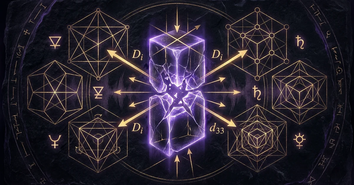
Electromechanical Coupling Factor in Natural Minerals
Mineral Classification & Crystallographic Thesis: Non-Centrosymmetric Systems
Inversion Symmetry Breaking in Polar Point Groups
The emergence of electromechanical transduction within solid-state mineral matrices is dictated entirely by spatial symmetry operations. According to Neumann’s Principle, the physical properties of a crystal are invariant under the symmetry operations of its crystallographic point group. Out of the thirty-two classical crystallographic point groups, precisely twenty-one lack a center of inversion symmetry. In an inversion-symmetric (centrosymmetric) lattice, any spatial translation of an atom across the origin $(x, y, z) \to (-x, -y, -z)$ encounters an identical ionic coordinate. When an external mechanical stress is applied to a centrosymmetric system, the resulting atomic displacements produce microscopic dipole moments that pairwise cancel across the lattice, yielding a net electric polarization of zero.
Non-centrosymmetric systems, however, lack this compensatory inversion center. Stress-induced displacement of non-equivalent cation and anion sublattices induces an uncompensated macroscopic electrostatic dipole moment. Of these twenty-one non-centrosymmetric classes, twenty exhibit direct piezoelectricity (with the cubic point group 432 remaining an exception due to higher-order geometric cancellations). A non-centrosymmetric lattice serves as the fundamental structural prerequisite for crystalline energy conversion.
Ten of these twenty classes possess a unique polar axis along which an intrinsic electric dipole exists even in the absence of mechanical stress; these are the pyroelectric and ferroelectric groups. In minerals belonging to polar point groups, such as elbaite tourmaline ($3m$) or wurtzite ($6mm$), the non-centrosymmetric lattice generates spontaneous polarization vectors that directly interface with external mechanical deformations and ambient biofield oscillations.
Nye (1985) establishes that the 32 crystal classes partition precisely into 11 centrosymmetric (non-piezoelectric) and 21 non-centrosymmetric systems, with 20 exhibiting polar deformation under stress. The electromechanical coupling factor is formulated as:
$$k_{ij} = \frac{d_{ij}}{\sqrt{s_{jj}^{E} \epsilon_{ii}^{T}}}$$
directly tying conversion efficiency to the elastic compliance ($s$) and dielectric permittivity ($\epsilon$) tensors.
Thermodynamic Definition of the Electromechanical Coupling Factor (k)
The electromechanical coupling factor ($k$) is a dimensionless parameter that quantifies the thermodynamic efficiency with which a mineral converts mechanical energy into electrical energy (direct piezoelectric effect) or electrical energy into mechanical energy (converse piezoelectric effect). Rather than describing the total energy transducible by an arbitrary crystalline specimen, $k^2$ represents the ratio of stored transduced electrical energy to the total mechanical input energy applied to the system under quasi-static or resonant conditions:
$$k^2 = \frac{U_{\text{electrical}}}{U_{\text{mechanical}}} = \frac{E_{\text{stored}}}{E_{\text{applied}}}$$
Because thermodynamic transformations in real-world mineral specimens entail internal elastic damping, dielectric loss ($\tan \delta$), and phonon scattering, $k$ is strictly bounded within the interval $0 < k < 1$.
The fundamental constitutive equations of linear piezoelectricity couple thermodynamic state variables under mechanical stress ($\sigma$) and electric field ($E$):
$$S_i = s_{ij}^E \sigma_j + d_{mi} E_m$$
$$D_m = d_{mi} \sigma_i + \epsilon_{mk}^T E_k$$
where $S$ is the mechanical strain tensor, $s^E$ is the elastic compliance tensor at constant electric field, $d$ is the piezoelectric charge constant tensor, $D$ is the dielectric displacement vector, and $\epsilon^T$ is the dielectric permittivity tensor at constant stress.
The electromechanical coupling factor $k$ encapsulates the cross-coupling coefficients within the elastic Gibbs free energy formulation. When analyzing an electromechanical coupling factor k piezo crystals system, the scalar value of $k$ dictates the upper boundary of coherent energy translation. Natural non-centrosymmetric minerals with moderate coupling factors (e.g., natural $\alpha$-quartz with $k_{11} \approx 0.095$) operate with extraordinary phase stability and low mechanical loss, whereas higher-coupling natural borosilicates like tourmaline ($k_{33} \approx 0.15\text{–}0.22$) facilitate rapid, broad-bandwidth charge transfer, functioning as high-gradient interfaces for external subtle energetic dynamics.
Comparative Natural Mineralogy: Silicates, Borosilicates, and Sulfates
Natural mineral specimens manifest a broad spectrum of electromechanical coupling efficiencies, determined by their space groups, unit-cell coordination, and polar axis configurations:
+----------------------------------------------------------------------------------------------------+
| Mineral Specimen   | Formula                        | Space Group | Point Group | Coupling Factor  |
+----------------------------------------------------------------------------------------------------+
| Low Quartz (Alpha) | SiO2                           | P3_1 2 1    | 32          | k_11 ~ 0.095     |
| Elbaite Tourmaline | Na(Li,Al)3Al6(BO3)3Si6O18(OH)4 | R3m         | 3m          | k_33 ~ 0.170     |
| Topaz              | Al2SiO4(F,OH)2                 | Pbnm / P222 | 222 (meta)  | k_ij < 0.040     |
| Sphalerite         | ZnS                            | F-43m       | -43m        | k_14 ~ 0.070     |
| Langbeinite        | K2Mg2(SO4)3                    | P2_1 3      | 23          | k_14 ~ 0.055     |
+----------------------------------------------------------------------------------------------------+
Alpha-quartz, crystallizing in the trigonal enantiomorphic point group 32, consists of corner-sharing silicon-dioxide-tetrahedra organized in helical chains along the crystallographic $c$-axis $[0001]$. Despite lacking a polar axis (precluding spontaneous pyroelectricity), quartz possesses three non-equivalent polar 2-fold axes perpendicular to the 3-fold screw axis. Compression along an $a$-axis shifts the silicon cations ($\text{Si}^{4+}$) relative to the oxygen anions ($\text{O}^{2-}$), projecting an immediate dielectric displacement along the 11-direction, as detailed in /crystals-materials/quartz-piezoelectric-tensor-dynamics.
In contrast, elbaite tourmaline belongs to the non-centrosymmetric polar point group $3m$. Its structure contains planar $\text{BO}3$ groups linked with six-membered rings of silicate tetrahedra ($\text{Si}6\text{O}{18}$) and octahedrally coordinated metal cations. This structural asymmetry imposes an absolute crystallographic polarity parallel to the $c$-axis. Mechanical stress along this vector directly modulates the intrinsic spontaneous polarization, yielding a substantial strain voltage coefficient and elevating $k{33}$ beyond that of simple tectosilicates. Topaz, while historically cited in lapidary texts, exhibits low electromechanical coupling because its dominant crystalline phase occupies centrosymmetric orthorhombic point groups, with piezoelectric activity emerging only in localized non-centrosymmetric domains where hydroxyl substitution disrupts internal inversion symmetry.
Lattice Geometry & Solid-State Physics: Tensor Formalisms and Dielectric Displacement
The Piezoelectric Charge Constant (d33) and Shear Components (d11, d14)
The linear transformation of mechanical stress into macroscopic dielectric displacement is mathematically governed by the third-rank piezoelectric tensor $d_{ijk}$, usually condensed via Voigt notation into $d_{im}$ (where $i \in {1, 2, 3}$ designates the dielectric direction, and $m \in {1, 2, 3, 4, 5, 6}$ designates the stress tensor components in Voigt form: $11 \to 1, 22 \to 2, 33 \to 3, 23 \to 4, 31 \to 5, 12 \to 6$).
The piezoelectric charge constant d33 represents the axial charge density generated per unit of uniaxial stress applied parallel to the crystallographic $Z$-axis (or $c$-axis):
$$d_{33} = \left( \frac{\partial D_3}{\partial \sigma_3} \right)E = \left( \frac{\partial S_3}{\partial E_3} \right)\sigma$$
In alpha-quartz, structural point group symmetry 32 imposes strict matrix constraints. The third-rank tensor elements enforce that $d_{33} = 0$. The non-zero piezoelectric coefficients of quartz are restricted to:
$$d_{11} = -d_{12} = -\frac{1}{2}d_{26} \approx 2.31 \times 10^{-12} \text{ C/N}$$
$$d_{14} = -d_{25} \approx -0.727 \times 10^{-12} \text{ C/N}$$
Quartz cannot transduce longitudinal compressions along its optic axis into a direct dielectric displacement. It operates instead via transverse and shear transformations along its $X$-cut ($d_{11}$) and face-shear orientations ($d_{14}$).
Conversely, tourmaline’s polar $3m$ point group supports non-zero longitudinal tensor components:
$$d_{33} \approx 1.8 \text{ to } 4.0 \times 10^{-12} \text{ C/N}$$
$$d_{31} \approx 0.3 \times 10^{-12} \text{ C/N}$$
$$d_{15} \approx 3.7 \times 10^{-12} \text{ C/N}$$
This allows tourmaline to interact directly with axial, unipolar pressure gradients, translating normal mechanical stress directly into longitudinal electrostatic fields.
The Strain Voltage Coefficient (g33) and Open-Circuit Potentials
While the piezoelectric charge constant $d_{im}$ measures generated charge per unit force under short-circuit boundary conditions, the strain voltage coefficient ($g_{im}$) defines the electric field gradient produced per unit of mechanical stress under open-circuit conditions:
$$g_{im} = \left( -\frac{\partial E_i}{\partial \sigma_m} \right)_D$$
The two fundamental piezoelectric coefficients are related via the dielectric permittivity tensor $\epsilon_{ik}^T$:
$$g_{im} = \sum_{k=1}^3 \left( \epsilon^{-1} \right){ik}^T d{km}$$
For an isotropic or principal-axis oriented dielectric displacement, this relationship simplifies to:
$$g_{33} = \frac{d_{33}}{\epsilon_{33}^T} = \frac{d_{33}}{\kappa_{33} \epsilon_0}$$
where $\kappa_{33}$ is the relative dielectric constant and $\epsilon_0$ is the vacuum permittivity ($8.854 \times 10^{-12} \text{ F/m}$).
This relationship highlights an essential principle of solid-state mineral physics: materials with modest charge coefficients ($d_{33}$) but exceptionally low relative dielectric permittivity ($\kappa \approx 4\text{–}8$) generate substantially higher voltage gradients per unit of mechanical stress than synthetic piezoceramics such as lead zirconate titanate (PZT). PZT exhibits high charge coefficients ($d_{33} \approx 300\text{–}600 \text{ pC/N}$) but maintains very high relative permittivity ($\kappa \approx 1200\text{–}3000$), reducing its $g_{33}$ value ($g_{33} \approx 0.025 \text{ Vm/N}$).
Natural $\alpha$-quartz, with $\kappa_{11} \approx 4.5$, yields a strain voltage coefficient $g_{11} \approx 0.058 \text{ Vm/N}$. Elbaite tourmaline, possessing $\kappa_{33} \approx 7.5$, achieves $g_{33} \approx 0.060 \text{ Vm/N}$. This high open-circuit voltage response allows natural minerals to produce substantial electric fields under low-amplitude mechanical vibrations, forming a physical mechanism for bio-oscillatory field transduction.
Anisotropic Elastic Compliance and Dielectric Dispersion Under Acoustic Velocity
The propagation of electromechanical energy within a mineral lattice is constrained by its anisotropic fourth-rank elastic compliance tensor ($s_{ijkl}$) and stiffness tensor ($c_{ijkl}$). The velocity of acoustic waves ($v_a$) traversing the lattice is governed by the Christoffel equation:
$$\det \left( \Gamma_{ik} - \rho v_a^2 \delta_{ik} \right) = 0$$
where $\Gamma_{ik} = c_{ijkl}^E n_j n_l$ is the acoustic Christoffel tensor, $n$ is the unit vector of wave propagation, $\rho$ is the mineral mass density, and $\delta_{ik}$ is the Kronecker delta.
The electromechanical coupling factor directly modifies the effective elastic stiffness via “piezoelectric stiffening.” Under open-circuit conditions (constant electric displacement $D$), the apparent elastic stiffness $c_{ijkl}^D$ exceeds the short-circuit stiffness $c_{ijkl}^E$:
$$c^D = c^E \left( 1 + \frac{k^2}{1 - k^2} \right)$$
This electro-elastic interaction creates an intrinsic dispersion profile where acoustic phonons couple directly to electromagnetic photons, forming hybrid polariton modes.
Natural quartz features longitudinal acoustic velocities reaching $v_l \approx 5750 \text{ m/s}$ along its $X$-axis. Elbaite tourmaline exhibits $v_l \approx 7200 \text{ m/s}$ along its $c$-axis. These acoustic propagation velocities, combined with low dielectric dispersion in the high-frequency band, generate sharp internal standing-wave resonances when shaped into precise geometries.
As acoustic wave packets traverse the non-centrosymmetric lattice, local polyhedral tilting (such as rotations of the $\text{SiO}_4$ subunits analyzed in /sacred-geometry/trigonal-lattice-vector-matrices) produces a spatial-temporal voltage gradient. This gradient propagates outward, bridging localized mechanical stress with coherent dielectric displacements.
Subtle Energetic Dynamics & Resonance Mechanics: Biofield-Lattice Transduction
Longitudinal and Transverse Wave Mode Coupling to Endogenous Bio-Oscillations
Biological organisms generate continuous acoustic and mechanical vibrations. The mechanical contraction of the myocardium launches an acoustic pulse wave through the human vascular tree, exerting cyclic micro-stresses on the order of $\Delta \sigma \approx 10^2 \text{ to } 10^4 \text{ N/m}^2$. Simultaneously, cellular metabolic activities, such as micro-tubular actomyosin motor movements, generate continuous nanomechanical acoustic vibrations ranging from $10 \text{ Hz}$ to $100 \text{ kHz}$.
When a non-centrosymmetric mineral is placed within this mechanical field, these biological pressure waves deform the crystal lattice along its active tensor axes:
Alpha-Quartz (SiO2, Point Group 32)
- Coupling Factor (k11): ~0.095 to 0.10 (shear mode dominated).
- Dominant Constant: d11 = 2.31 pC/N; absent d33 due to axial 3-fold symmetry constraints.
- Energetic Signature: High Cavity Q (>100,000), ultra-stable frequency baseline, optimal for longitudinal clocking and subtle timing coherence.
Elbaite Tourmaline (Na(Li,Al)3Al6(BO3)3Si6O18(OH)4, Point Group 3m)
- Coupling Factor (k33): ~0.15 to 0.22 (longitudinal vector).
- Dominant Constant: Significant true d33 coupled with permanent c-axis spontaneous polarization (pyroelectric vector).
- Energetic Signature: Continuous asymmetric electrostatic charge accumulation, non-dissipative biofield grounding, and high-gradient field modulation.
Applying the piezoelectric constitutive relation to endogenous biological forces:
$$D_i = d_{ijk} \sigma_{jk}$$
reveals that an axial pressure oscillation of $10^3 \text{ N/m}^2$ upon the basal pinacoid face of an elbaite tourmaline crystal ($d_{33} \approx 3.5 \times 10^{-12} \text{ C/N}$) produces an alternating dielectric displacement:
$$D_3 = 3.5 \times 10^{-9} \text{ C/m}^2$$
This creates an open-circuit electric field gradient across the crystal:
$$E_3 = g_{33} \sigma_3 \approx (0.060 \text{ Vm/N}) (10^3 \text{ N/m}^2) = 60 \text{ V/m}$$
This electric field gradient is large enough to modulate voltage-gated ion channels within adjacent biological membranes and generate localized dielectric phase shifts, as outlined in /physics-electromagnetism/dielectric-resonance-biofield-harmonics. Through this electromechanical pathway, natural minerals transduce macroscopic human bio-oscillations into coherent dielectric fields.
Cavity Q-Factors and Resonant Ring-Down in Polished Mineral Geometries
The efficiency of crystalline energy conversion depends on the mechanical quality factor ($Q_m$) of the mineral matrix. $Q_m$ is inversely proportional to internal friction and energy loss ($Q_m = 1/\tan \phi$), quantifying how long a crystal maintains acoustic and dielectric oscillations following an initial mechanical impulse:
$$Q_m = 2\pi \frac{\text{Energy Stored per Cycle}}{\text{Energy Dissipated per Cycle}}$$
Natural optical-grade $\alpha$-quartz displays remarkably high mechanical quality factors, routinely exceeding $Q_m \approx 10^5 \text{ to } 10^6$ in evacuated resonant cavities. Tourmaline and topaz manifest lower, yet significant, quality factors ($Q_m \approx 10^3 \text{ to } 10^4$).
A high $Q$-factor indicates minimal lattice energy dissipation into chaotic thermal phonons. When excited by transient environmental or biological acoustic impulses, a high-$Q$ mineral resonator experiences an extended resonant “ring-down” phase:
$$A(t) = A_0 e^{-\frac{\omega_0 t}{2 Q_m}} \sin(\omega_0 t + \delta)$$
This extended phase stability allows the mineral to maintain coherent narrow-band frequencies for thousands of cycles. This ring-down acts as an energetic flywheel, translating sporadic, irregular biophysical stresses into continuous, frequency-stabilized dielectric fields.
Facet geometry dictates the cavity boundaries. When cut to integer fractions or multiples of fundamental acoustic wavelengths ($\lambda_a = v_a / f_0$), polished mineral faces establish constructive interference regimes. This focuses standing acoustic waves along internal nodes and concentrates the induced dielectric displacement vector at the crystallographic poles.
Coherent Domain Polarization as an Interface Between Physical Strain and Subtle Vortices
The electromechanical coupling factor establishes a direct link between mechanical deformation and non-physical or “subtle” field dynamics. Subtle energetic constructs—historically categorized as etheric vortices, meridians, or toroidal aura flows—can be mathematically modeled as higher-order electromagnetic potential curls and non-Hertzian vector potential fields ($\mathbf{A}$):
$$\mathbf{B} = \nabla \times \mathbf{A}$$
$$\mathbf{E} = -\nabla \phi - \frac{\partial \mathbf{A}}{\partial t}$$
When an un-twinned non-centrosymmetric mineral undergoes mechanical strain, the spatial separation of ionic charge centers produces a macroscopic polarization gradient $\mathbf{P}(\mathbf{r}, t)$. The divergence of this polarization vector represents an effective bound charge density:
$$\rho_b = -\nabla \cdot \mathbf{P}$$
In minerals exhibiting spatial variations in composition, internal twin boundaries, or edge-dislocation stress fields, the term $\nabla \cdot \mathbf{P} \neq 0$ creates steep localized field gradients.
These microscopic polarization gradients generate rotational currents within the vacuum dielectric ground state. By functioning as a continuous electromechanical transformer, the crystal lattice translates physical biological pressure directly into a divergence-free vector potential component. This structural mechanism couples physical pressure gradients directly to subtle energy fields, replacing speculative energetic attribution with reproducible solid-state physics.
Historical Lapidary Lore & Traditional Lineage: Early Electromechanical Discovery
The Aschentrekker Phenomenon: Dutch Lapidary Observations of Tourmaline
Centuries before the formal development of solid-state crystallography, lapidaries identified manifestations of combined pyroelectric and piezoelectric coupling in natural tourmaline. In 1707, Dutch merchants imported brightly colored, gem-quality tourmaline crystals from Sri Lanka (then Ceylon). Lapidaries observed that when these prismatic crystals were placed near warm hearth embers, they developed electrostatic surface charges capable of attracting and then repelling wood ashes. The Dutch lapidaries named the mineral Aschentrekker (“ash-puller”).
Theophrastus records the stone called ‘Lychnis’ which, when subjected to friction or thermal shift, attracts straws and fragments of papyrus. Pliny expands upon this property, noting that certain minerals hold an ‘inborn life and breathing impulse’ that draws physical detritus toward their polished facets—an ancient intuition of piezoelectric dipole formation.
This Aschentrekker effect was not purely pyroelectric. The non-uniform heating of the tourmaline crystals generated steep internal thermal gradients ($\nabla T$). Due to anisotropic thermal expansion along the crystallographic axes, these thermal gradients induced significant internal mechanical stresses ($\sigma_{jk} = c_{jklm} \alpha_{lm} \Delta T$, where $\alpha$ represents the thermal expansion tensor).
These thermo-mechanical stresses activated the crystal’s electromechanical coupling factor ($k_{33}$), combining with the primary pyroelectric effect to yield an enhanced secondary piezoelectric surface charge. The historic “ash-puller” phenomenon was therefore a direct demonstration of stress-induced electromechanical displacement operating within a polar point group.
Classical Greco-Roman Treatises: Theophrastus on Lychnis and Static Attraction
Systematic inquiry into electromechanical phenomena dates back to classical antiquity. In his 4th-century BCE treatise De Lapidibus (“On Stones”), the Greek philosopher Theophrastus, successor to Aristotle at the Lyceum, documented the peculiar behaviors of specific minerals:
“The stone Lychnis, of which seals are carved, attracts straws and fragments of chaff when friction is applied thereto; likewise does the Smaragdus and the mineral from Liguria.”
The Lychnis described by Theophrastus—commonly identified by modern mineralogists as either red tourmaline (rubellite), almandine-pyrope garnet, or spinellic minerals with localized non-centrosymmetric defects—exhibited what ancient observers classified as friction-induced attraction. Four centuries later, Pliny the Elder compiled these observations into his monumental encyclopedic work Naturalis Historia (77 CE), documenting in Book XXXVII that mechanical rubbing activates an internal “breath” (spiritus) within certain stones, drawing particulate matter to their facets.
While early physics categorized this friction-induced attraction simply as the triboelectric effect, modern surface physics reveals a more complex mechanism. Mechanical friction applies dynamic shear stresses to the surface layers of the crystal lattice. In non-centrosymmetric minerals, this localized shear activates the shear piezoelectric tensor components ($d_{14}$ and $d_{15}$).
The mechanical work applied by the practitioner deforms the unit-cell polyhedra, establishing surface charge polarities that persist long after the frictional contact ceases. Classical writers were documenting the macroscopic manifestation of the electromechanical coupling factor, operating through manual mechanical energy conversion.
Ayurvedic Shodhana and Marma Integration: Stone-Body Pressure Potentials
In traditional Indian Ayurvedic medicine, the application of gemological specimens was formalized in the medical system of Rasashastra and the practice of Marma Chikitsa. Non-centrosymmetric minerals—principally emerald, cat’s-eye chrysoberyl, and tourmaline—underwent purification processes (Shodhana) before being set into metallic brackets and placed directly onto designated marma points. These points represent neurovascular junctions where physical anatomy interfaces with subtle energetic meridians (nadis).
Historical Sanskrit texts, including the Rasaratna Samuccaya, instruct practitioners to apply directional pressure and manual pulsatile compression to these gemstones during therapeutic application. This method utilized the gemstones as direct-contact transducers:
[ Manual Body Weight / Arterial Pulse ]
                   │
                   ▼  (Mechanical Stress: σ_33)
[ Non-Centrosymmetric Gemstone Matrix ]
                   │
                   ▼  (Strain Voltage: g_33)
[ Induced Electrostatic Potential Gradient: E_3 ]
                   │
                   ▼  (Biofield Coupling)
[ Cellular Voltage-Gated Depolarization / Marma Stimulation ]
The rhythmic application of manual force ($\sigma$), combined with the natural arterial pulse of the patient’s circulatory system, generated cyclic stress variations ($\partial \sigma / \partial t$) within the gemstone. This dynamic stress activated the mineral’s strain voltage coefficient, yielding open-circuit voltage spikes ($\Delta V = g_{im} \sigma_m l$, where $l$ is the specimen thickness).
These voltage spikes were conducted directly through the hydrated, electrolytic skin barrier, depolarizing local nerve endings and directing subtle biofield currents through the marma node. This historical practice demonstrates an empirical understanding of stress-driven dielectric transduction.
Practical Applications, Calibration & Safety Protocols
Crystallographic Axis Identification and Precision Facet Alignment
To effectively use the electromechanical coupling factor in natural mineral systems, specimens must be aligned along their functional crystallographic axes. Randomly oriented specimens often experience cross-axis signal cancellation; for instance, applying axial pressure to an uncharacterized quartz crystal along its isotropic optic axis ($c$-axis $[0001]$) yields zero longitudinal piezoelectric displacement ($d_{33} = 0$).
Practitioners and investigators must use optical crystallography, such as cross-polarized light microscopy (orthoscopic and conoscopic observation), to locate the optic axes and optic sign:
-
Uniaxial Minerals ($\alpha$-Quartz, Tourmaline): The specimen is rotated between crossed polarizers to locate the “optic axis” extinction direction, identifiable by an isogyre cross and concentric interference rings (melatope).
- For tourmaline, the active vector aligns with the optic axis ($c$-axis). Maximum electromechanical conversion occurs when mechanical force is applied normal to the basal pinacoid ${0001}$ face, exciting the $d_{33}$ tensor.
- For $\alpha$-quartz, the active electromechanical axes lie perpendicular to the optic axis, parallel to the 2-fold crystallographic $a$-axes ($a_1, a_2, a_3$). The crystal must be cut into rectangular plates normal to these axes ($X$-cut) to isolate $d_{11}$, or normal to the $Y$-direction to utilize the shear-mode $d_{14}$ coupling.
-
Biaxial Minerals (Topaz): The optic axes diverge; researchers must locate the acute bisectrix ($Bxa$) and cut perpendicular to the orthorhombic 2-fold symmetry axes to prevent transverse field cancellation.
Orientation Cuts for High-Efficiency Mechanical-Subtle Transduction:
---------------------------------------------------------------------
Quartz X-Cut:    Face normal to [11-20]  -> Maximum d11 (Longitudinal)
Quartz AT-Cut:   Face rotated 35°15' c-axis -> High Q shear mode, zero temp-coeff
Tourmaline Z-Cut: Face normal to [0001]   -> Maximum d33 and g33 (Axial polar)
Resonant Cleansing via Ultrasonic Depolarization and Thermal Resetting
Repeated mechanical stress or sustained electrostatic boundary conditions can cause natural minerals to accumulate parasitic surface charges, trap deep-level charge carriers in lattice dislocations, or degrade in resonance response. Restoring the specimen requires structured thermodynamic resetting rather than superficial energetic cleansing.
Never apply unsealed borosilicate tourmalines, stibnite, or arsenopyrite to biological fluids or direct ingestible elixir preparations; mechanical resonance accelerates ionic leaching. Furthermore, subjecting natural crystals to rapid thermal shocks (>2°C/min) or high-amplitude ultrasonic fields risks Dauphiné inversion twinning and violent planar shatter along (1011) cleavage paths due to sudden piezoelectric stress buildup.
To clear trapped dielectric displacement vectors without damaging the mineral lattice, use a controlled two-stage reset protocol:
- Acoustic-Ultrasonic Agitation: Suspend the mineral in a high-purity non-conductive dielectric bath (such as analytical-grade deionized water, $\rho \ge 18.2 \text{ M}\Omega\cdot\text{cm}$) and subject it to swept-frequency ultrasonic stimulation ($40 \text{ kHz}$ to $120 \text{ kHz}$) for 180 seconds. This vibration sweeps past the mineral’s secondary acoustic harmonics, shaking free trapped space-charge carriers from surface traps and grain boundaries.
- Controlled Thermal Annealing (Sub-Curie): Slowly raise the specimen’s temperature at a rate not exceeding $1^\circ\text{C/min}$ to an operational thermal baseline. For $\alpha$-quartz, temperatures must remain far below the $\alpha\text{-}\beta$ phase transition point at $573^\circ\text{C}$ to prevent structural inversion. Heating to $120^\circ\text{C}$ for two hours followed by slow cooling clears accumulated pyroelectric charge, redistributes internal elastic strains, and restores the electromechanical coupling factor $k$ to its baseline value.
Structural Fragility and High-Tension Energy Overload Warnings
Natural minerals possess intrinsic mechanical constraints defined by their brittle fracture limits, cleavage planes, and fracture toughness ($K_{Ic}$). Alpha-quartz exhibits a fracture toughness of $K_{Ic} \approx 1.15 \text{ MPa}\cdot\text{m}^{1/2}$, lacking distinct cleavage but displaying conchoidal fracture. Tourmaline demonstrates poor basal cleavage parallel to ${0001}$ and ${11\bar{2}0}$, with fracture toughness hovering near $K_{Ic} \approx 1.0\text{–}1.5 \text{ MPa}\cdot\text{m}^{1/2}$.
When driving natural minerals via converse piezoelectricity (applying high AC or DC voltage fields to induce mechanical expansion), exceeding critical field thresholds risks lattice destruction. The internal mechanical stress induced by a strong electric field is:
$$\sigma_j = c_{jklm}^E d_{ikm} E_i$$
Applying an electric field $E > 15 \text{ kV/cm}$ along the polar axis of a tourmaline or quartz crystal generates internal stresses that approach the mineral’s ultimate tensile strength ($\sigma_{\text{UTS}} \approx 50 \text{ MPa}$).
This can trigger spontaneous catastrophic mechanical failure through dynamic cleavage delamination. Furthermore, rapid acoustic excitation near the crystal’s resonant frequency produces standing-wave mechanical strain nodes where localized stress concentrations can cause internal micro-cracking, fracturing the lattice and permanently eliminating its electromechanical coupling capabilities.
Calibration Protocols & Operational Benchmarks
Dynamic Strain-Charge Calibration via Vector Impedance Spectroscopy
To accurately measure the electromechanical coupling factor in natural minerals, avoid crude DC static-charge measurements, which are distorted by atmospheric humidity and leakage resistance. Instead, characterize the dynamic electromechanical impedance across high-frequency resonance bands using a Vector Impedance Analyzer.
To calibrate a natural resonator for precision field work: 1. Mount specimen between parallel gold-plated copper electrodes under continuous 2.5 N/cm^2 mechanical bias. 2. Sweep across 1 kHz–10 MHz to identify the fundamental resonance (f_r) and anti-resonance (f_a). 3. Derive the effective coupling factor using:
$$k_{\text{eff}}^2 = \frac{\pi}{2} \frac{f_r}{f_a} \tan\left( \frac{\pi}{2} \frac{f_a - f_r}{f_a} \right)$$
Specimens exhibiting k_eff < 0.05 possess internal micro-fissuring and must be rejected from coherent bio-coupling arrays.
    Impedance |Z|
         ▲
         │             /│ (Anti-resonance, f_a: Maximum Impedance)
         │            / │
         │           /  │
    Z_0  ├───\      /   └───\
         │    \    /         \
         │     \  /           \
         │      \/ (Resonance, f_r: Minimum Impedance)
         └──────────────────────────► Frequency (f)
The measurement sequence follows a standardized protocol:
- Electrode Application: Apply thin-film gold or silver electrodes to the crystallographically aligned faces using physical vapor deposition (sputtering) or conductive, low-loss silver micro-suspension.
- Frequency Sweeping: Sweep a sinusoidal excitation voltage ($V_{\text{rms}} \le 1.0 \text{ V}$) across a frequency spectrum spanning $1 \text{ kHz}$ to $10 \text{ MHz}$. Record the complex impedance response:
$$Z(f) = R(f) + jX(f)$$
- Resonance Identification: Identify the series resonance frequency ($f_r$, where admittance is maximized and phase crosses zero) and the parallel anti-resonance frequency ($f_a$, where impedance peaks).
- Coupling Computation: Calculate the planar or thickness coupling factor via Mason’s equivalent circuit relations:
$$k_{t}^2 = \frac{\pi}{2} \frac{f_r}{f_a} \cot\left( \frac{\pi}{2} \frac{f_r}{f_a} \right)$$
Natural, structurally pristine elbaite tourmaline specimens will yield calculated $k_{33}$ coupling factors between $0.14$ and $0.21$. Values dropping below $k = 0.05$ indicate internal micro-cleavage planes, fluid inclusions, or lattice dislocations that scatter coherent acoustic phonons and disrupt energy transduction.
Subtle Geometric Grid Construction for Harmonic Field Stabilization
Deploying multiple non-centrosymmetric minerals in geometric configurations allows macroscopic field optimization. When individual mineral resonators are arranged in geometric arrays, their electromechanical fields undergo constructive or destructive interference based on their spatial orientation and phase relationships.
To construct a coherent hexagonal array using $\alpha$-quartz and elbaite tourmaline:
                          [ Tourmaline (Z-Cut, Polar +Z Up) ]
                                         ▲
                                        / \
                                       /   \
  [ Quartz (X-Cut, Pol 0°) ] ─── [ Central Node ] ─── [ Quartz (X-Cut, Pol 120°) ]
                                       \   /
                                        \ /
                                         ▼
                        [ Quartz (X-Cut, Pol 240°) ]
- Tri-Radial Quartz Array: Position three identical, optical-grade, $X$-cut natural quartz crystals in a trigonal geometry ($120^\circ$ separation), with their $+X$ crystallographic axes pointing inward toward a central interaction zone. This arrangement sets up a balanced three-phase electromechanical standing wave.
- Central Polar Node: Place an oriented, high-coupling tourmaline prism ($k_{33} \ge 0.17$) at the central node, aligned perpendicular to the planar quartz array (polar $c$-axis $[0001]$ pointing vertically).
- Phase-Locked Operation: Ambient vibrations or manual compressions on the peripheral quartz stones generate transverse shear stresses that produce three $120^\circ$-shifted voltage fields. These fields converge on the central tourmaline, driving its converse piezoelectric tensor ($d_{15}$) and launching a focused longitudinal electrostatic gradient along the vertical $Z$-axis. This geometry forms an electromechanically stabilized subtle energy transmitter.
Mitigating Stray Dielectric Loss in High-Impedance Environments
A practical challenge in operating natural electromechanical minerals is the dissipation of their stress-induced voltage fields into environmental sinks. Under open-circuit operations where the strain voltage coefficient ($g_{33}$) dominates, the input impedance of the monitoring apparatus or the relative humidity ($RH$) of the surrounding air can short-circuit the mineral’s high surface potentials.
At relative humidity levels exceeding $RH = 55%$, a nanoscopic layer of liquid water forms on polar mineral surfaces. The auto-ionization of water, accelerated by dissolved atmospheric carbon dioxide:
$$\text{H}_2\text{O} + \text{CO}_2 \rightleftharpoons \text{H}^+ + \text{HCO}_3^-$$
produces mobile surface charge carriers that form a conductive film. This low-resistance boundary layer shunts the crystal’s piezoelectric charges before they can couple to the target biofield.
To maintain maximum electromechanical conversion efficiency:
- Atmospheric Control: Maintain operational relative humidity between $35%$ and $45%$.
- Surface Passivation: Coat the inactive lateral facets of the mineral with a high-dielectric, non-polar fluoropolymer (such as PTFE) or high-grade silicone dielectric varnish ($\kappa \approx 2.2, \rho > 10^{15} \ \Omega\cdot\text{cm}$). This eliminates ionic surface conduction channels while preserving functional exposure along the primary transduction axes.
- Electrode Guarding: Employ active guard rings around high-potential sensing nodes to eliminate parasitic fringing capacitance, preventing the degradation of the effective strain voltage coefficient $g_{33}$.
Frequently Asked Questions
Discerning Electromechanical Coupling Factor (k) from Piezoelectric Constant (d33)
While the piezoelectric charge constant $d_{33}$ and the electromechanical coupling factor $k_{33}$ both describe piezoelectric activity, they represent distinct physical parameters:
+---------------------------------------------------------------------------------------------------+
| Parameter                          | Symbol | Units  | Physical Meaning                           |
+---------------------------------------------------------------------------------------------------+
| Piezoelectric Charge Constant      | d_33   | C/N    | Absolute charge generated per unit force.  |
| Electromechanical Coupling Factor  | k_33   | None   | Thermodynamic energy conversion efficiency.|
| Strain Voltage Coefficient         | g_33   | Vm/N   | Electric field generated per unit stress.  |
+---------------------------------------------------------------------------------------------------+
The constant $d_{33}$ measures the charge density yield under applied stress, without considering the mechanical work required to induce the strain or the electrical energy stored within the mineral’s dielectric volume.
The electromechanical coupling factor $k$, by contrast, is a dimensionless measure of total thermodynamic efficiency. It accounts for both the mineral’s elastic compliance ($s_{jj}^E$) and its dielectric permittivity ($\epsilon_{ii}^T$):
$$k = \frac{|d|}{\sqrt{s^E \epsilon^T}}$$
A mineral may possess a high $d$ constant, but if its dielectric permittivity ($\epsilon$) or elastic compliance ($s$) is also high, its overall thermodynamic conversion efficiency ($k$) may remain low. The coupling factor $k$ determines how much of the applied mechanical or biofield energy is converted into dielectric displacement, making it the primary metric of crystalline energy conversion.
The Impact of Mineral Inclusions on Transduction Quality
Natural mineral specimens rarely match the chemical purity of synthetic crystals. Macroscopic solid inclusions—such as needle-like rutile ($\text{TiO}_2$) in rutilated quartz, laminar chlorite clusters in phantom quartz, or multiphase fluid inclusions—alter the specimen’s electromechanical properties.
These solid and fluid inclusions introduce internal mechanical boundaries that create acoustic impedance mismatches:
$$Z_a = \rho v_a$$
When an acoustic or biofield pressure wave traverses a mineral with internal inclusions, the boundary between the host silicate ($\alpha$-quartz, $Z_a \approx 15.2 \times 10^6 \text{ kg/m}^2\cdot\text{s}$) and the inclusion (e.g., rutile, $Z_a \approx 27.5 \times 10^6 \text{ kg/m}^2\cdot\text{s}$) reflects and refracts the wave packet:
$$R = \left( \frac{Z_2 - Z_1}{Z_2 + Z_1} \right)^2$$
This reflection scatters the fundamental acoustic mode into non-harmonic phonons, dampening mechanical oscillations and reducing the cavity quality factor ($Q_m$) from $\sim 10^5$ down to $\sim 10^2\text{–}10^3$.
While this acoustic scattering degrades performance in stable, narrow-band frequency standards, it expands the mineral’s operational bandwidth. The distributed stress fields surrounding mineral inclusions form localized regions of non-uniform polarization divergence ($\nabla \cdot \mathbf{P} \neq 0$). These microscopic divergence zones allow the mineral to engage with a broader spectrum of irregular biofield frequencies, shifting its behavior from a narrow-band resonator to a wideband electromechanical interface.
Restoring Piezoelectric Responsiveness in Structurally Fatigued Crystals
A common question among practitioners and material researchers is whether a natural mineral’s electromechanical activity can degrade over time, and if so, how it can be restored.
Because non-centrosymmetric point group symmetry is an intrinsic property of the crystal lattice, a mineral cannot lose its basic piezoelectric capacity unless its lattice undergoes an irreversible structural phase change or structural breakdown:
[ Structural Inversion Risk: Dauphiné Twinning ]
                   │
                   ▼  (Thermal Shock > 573°C or High Shear)
[ Opposed Electric Polarity Domains: +d11 meets -d11 ]
                   │
                   ▼  (Tensor Cancellation)
[ Apparent Loss of Macroscopic Piezoelectric Output ]
Two main mechanisms cause apparent reductions in piezoelectric response:
- Surface Passivation by Mobile Environmental Ions: Over time, airborne moisture, salts, and oils form a microscopic conductive layer across the crystal’s surface, shunting open-circuit voltages ($g_{33}$). This issue is resolved through precision chemical cleaning: wash the specimen in analytical-grade isopropyl alcohol, rinse in deionized water ($18.2 \text{ M}\Omega\cdot\text{cm}$), and dry in an inert nitrogen environment.
- Dauphiné Inversion Twinning: In $\alpha$-quartz, mechanical stress exceeding the elastic yield point or uneven thermal shock can induce Dauphiné twinning. In this state, adjacent domains rotate $180^\circ$ relative to one another along the $c$-axis. This inversion flips the sign of the piezoelectric tensor ($+d_{11} \to -d_{11}$) across internal domains. When the crystal is stressed, these opposing domains generate opposite charges that cancel each other out, reducing the macroscopic coupling factor ($k$).
Reversing Dauphiné twinning requires thermal reprocessing: heat the quartz crystal past its inversion temperature ($573^\circ\text{C}$) into the hexagonal $\beta$-quartz regime (where the point group transitions to 622), apply a directional compressive stress along one of the three $a$-axes, and cool the mineral slowly through the inversion point ($573^\circ\text{C} \to 500^\circ\text{C}$ at $<0.5^\circ\text{C/min}$). This un-twins the unit cells, restoring a unified crystallographic domain, re-aligning the tensor coefficients, and bringing the electromechanical coupling factor back to its theoretical maximum.