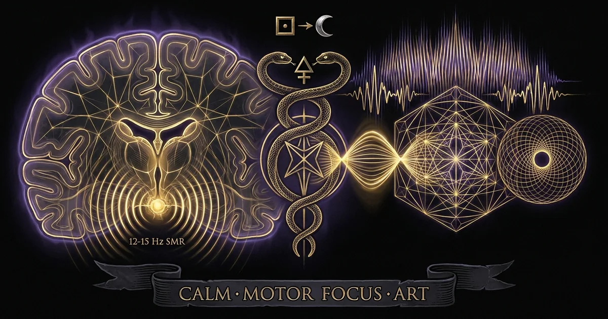
Sensorimotor Rhythm SMR (12-15 Hz): Calm Motor Focus Art
Protocol Overview & Neurophysiological Thesis: The Rolandic Idling Rhythm
The sensorimotor rhythm (SMR) designates an electroencephalographic oscillatory pattern localized within the 12–15 Hz band, captured prominently across the central rolandic cortex overlying the precentral and postcentral gyri. Functionally, SMR denotes a specialized thalamocortical idling state: it emerges spontaneously during wakeful, motionless alertness and attenuates immediately upon either the actual execution or the covert mental imagery of somatosensory processing and somatomotor output. Operant up-regulation of this rhythm enables targeted somatic stabilization, systematically decoupling attentional focus from peripheral neuromuscular drive. This forms an empirical bridge connecting clinical neuroregulation to classical contemplative stillness.
                  ┌───────────────────────────────┐
                  │      CORTICAL TOPOGRAPHY      │
                  │   Pre- & Post-Central Gyri    │
                  │       (Cz, C3, and C4)        │
                  └───────────────┬───────────────┘
                                  │
                                  ▼
┌─────────────────────────────────┴─────────────────────────────────┐
│              VENTROBASAL THALAMOCORTICAL PACEMAKER                │
│ 1. Ventrobasal Complex   ──> Somatosensory Inflow Gated           │
│ 2. Reticular Nucleus     ──> GABAergic Hyperpolarization (12-15Hz)│
│ 3. Ventral Lateral / Ant ──> Corticospinal Attenuation            │
└─────────────────────────────────┬─────────────────────────────────┘
                                  │
                                  ▼
┌─────────────────────────────────┴─────────────────────────────────┐
│                     SYSTEMIC SOMATIC OUTCOME                      │
│   • Corticomotor De-efference (Suppression of Peripheral Jitter)   │
│   • Autonomic Stabilization (Increased Parasympathetic HRV)       │
│   • Alert Cognitive Quiescence (Mind Awake, Somatomotor Still)   │
└───────────────────────────────────────────────────────────────────┘
Cytoarchitecture of the Sensorimotor Strip (Cz, C3, C4)
The primary electrophysiological nexus for SMR recording rests at the 10–20 international system coordinates corresponding to Cz, C3, and C4, situated immediately adjacent to the central sulcus (the Rolandic fissure). Cytoarchitectonically, this topographical zone encompasses Brodmann Area 4 (the primary motor cortex, M1) on the precentral bank and Brodmann Areas 3, 1, and 2 (the primary somatosensory cortex, S1) within the postcentral gyrus. M1 is marked by large pyramidal neurons within cortical layer V—most notably the giant cells of Betz—whose axons project down through the internal capsule, forming the corticospinal tract that governs voluntary peripheral musculature.
Directly contiguous across the sulcus, S1 exhibits a pronounced granular layer IV specialized in decoding primary somatic afferents arriving from peripheral mechanoreceptors, proprioceptive spindle fibers, and joint receptors. When an individual achieves deliberate, complete motor quiescence without falling into somnolence, the cross-talk between the primary motor efferent generators of Area 4 and the primary somatosensory afferent recipients of Areas 3, 1, and 2 drops below operational thresholds. This precise anatomical de-excitation manifests over the scalp electrodes at Cz (representing the bilateral lower extremities and core somatotopy along the medial longitudinal fissure), C3, and C4 (representing the contralateral upper extremities, hands, and facial regions) as a localized, high-amplitude spindle-burst phenomenon: the classical sensorimotor rhythm smr neurofeedback motor stillness signature.
Thalamocortical Somatosensory Gating and Ventrobasal Synchrony
The neurogenesis of SMR does not originate within the neocortex itself; rather, it reflects synchronized feedback within the thalamocortical loop, explicitly governed by the ventrobasal (VB) thalamic nuclear complex and the reticular thalamic nucleus (TRN). The ventrobasal complex acts as the obligatory relay station for the medial lemniscal pathway, which transmits deep tactile and conscious proprioceptive data from the spinal cord to the cerebral cortex. Encapsulating this thalamic core is the TRN, an exclusively GABAergic shell that monitors and modifies both ascending sensory information destined for the neocortex and descending corticothalamic projections returning from layer VI pyramidal cells.
Under conditions of active motor interrogation or physical agitation, sensory afferents barrage the ventrobasal complex, driving individual relay neurons into rapid single-spike transmission modes that correspond to low-amplitude, desynchronized beta activity (15–30 Hz) on the surface EEG. When somatic stillness is established, afferent signaling through the dorsal columns abates. Deprived of disruptive peripheral drive, TRN neurons switch their firing configuration into rhythmic burst-firing patterns mediated by low-threshold, T-type voltage-gated calcium channels ($Ca_v3.1, Ca_v3.2$). This rhythmic burst firing imposes alternating intervals of prolonged hyperpolarization and rebound depolarization upon the ventrobasal and ventral lateral thalamic nuclei. Consequently, ascending transmission to M1 and S1 is periodically clamped. The resulting synchronized rhythmic burst projected to the sensorimotor cortex establishes the 12–15 Hz oscillatory baseline characteristic of the idling sensorimotor strip.
Operant Conditioning of Endogenous Motor Stillness
The voluntary enhancement of the 12–15 Hz band constitutes one of the most reliable and thoroughly validated targets within clinical biofeedback. Operant conditioning leverages neuroplasticity to structurally calibrate the underlying thalamocortical pacemakers through real-time informational reinforcement. When a subject receives immediate auditory or visual rewards whenever their sensorimotor strip power within the 12–15 Hz window exceeds a designated threshold—while simultaneously penalizing theta (4–8 Hz) and high-beta (20–30 Hz) bands—the central nervous system optimizes its internal state to satisfy the contingency.
Mechanistically, this operant training directly suppresses involuntary somatic tremor, hyperkinetic neuromuscular discharges, and peripheral muscular tension. Because SMR synchronization requires the dampening of motor commands originating in Betz cells, sustained operant reinforcement shifts the central motor threshold, demanding significantly greater depolarizing current before corticospinal efferent bursts can trigger voluntary movement. This endogenous up-regulation directly translates into profound behavioral immobility, dampening the motor unrest characteristic of attention-deficit profiles while shielding the cortical architecture from paroxysmal epileptogenic discharges.
Sterman, M. B., & Friar, L. (1972). Suppression of seizures in an epileptic following sensorimotor EEG feedback training. Electroencephalography and Clinical Neurophysiology, 33(1), 89–95. This landmark investigation validated the translational application of feline operant SMR conditioning to human clinical pathology. Following initial aerospace trials demonstrating that feline subjects trained to produce SMR exhibited markedly elevated thresholds against monomethylhydrazine-induced toxicity and tonic-clonic convulsions, Sterman and Friar trained a medically refractory human epileptic patient. Operant reinforcement of the central sulcus 12–15 Hz rhythm yielded dramatic reductions in both the frequency and amplitude of major motor seizures, demonstrating that voluntary sensorimotor stabilization structurally alters baseline corticospinal excitability.
Biophysical Mechanisms & Brainwave Dynamics: Thalamocortical Pacemaking and Somatic De-efference
Understanding the biophysical operations underlying the 12–15 Hz band requires a detailed analysis of thalamocortical microcircuitry, particularly regarding how inhibitory rhythms decouple attentional vigilance from peripheral motor execution. The rhythm represents a unique physiological configuration: it is neither an active processing frequency like fast beta nor an internally absorbing, hypnagogic state like deep theta. It is a state of motor de-efference characterized by high internal alertness paired with systemic physical disengagement, which can be contextualized within broader frameworks of EEG frequency bands and thalamic gating.
        Afferent Proprioceptive Dampening
                      │
                      ▼
         TRN GABAergic Hyperpolarization
       (T-type Ca²⁺ Burst Mode: 12-15 Hz)
                      │
                      ▼
     Ventrobasal & Ventral Lateral Thalamic Gating
                      │
                      ▼
        Primary Sensorimotor Strip (Cz)
    Synchronous Burst Idling / Postsynaptic IPSPs
                      │
                      ▼
      Corticospinal Tract Volley Attenuation
  ┌───────────────────┴───────────────────┐
  ▼                                       ▼
Neuromuscular System:           Ascending Reticular Drive:
Absolute Skeletal Stillness     Sustained Cortical Vigilance
The Ventrobasal Complex and Reticular Thalamic Nucleus (TRN) Bursting
The cellular engine responsible for sensorimotor rhythm synchrony resides in the reciprocal synaptic loop connecting the reticular thalamic nucleus (TRN) with the specific relay neurons of the ventrobasal (VB) thalamus. The TRN is structurally positioned to intercept every reciprocal axon traveling between the neocortex and the deeper thalamic nuclei. When peripheral motor activity diminishes, the background tonic depolarization driven by the ascending reticular activating system (ARAS) undergoes localized modulation: cholinergic and noradrenergic afferents that favor single-spike tonic firing retreat, allowing TRN cells to hyperpolarize toward a resting membrane potential near -65 to -70 mV.
At this hyperpolarized potential, the inactivation gates of T-type low-threshold calcium channels deinactivate. When an excitatory corticothalamic or intrathalamic input impinges upon these primed TRN cells, it triggers low-threshold calcium spikes ($LTCS$), producing high-frequency bursts of action potentials at 200–400 Hz. These TRN bursts release large quantities of gamma-aminobutyric acid (GABA) onto the post-synaptic receptors of the ventrobasal thalamocortical relay cells. This synaptic event generates profound inhibitory postsynaptic potentials (IPSPs), transiently silencing the relay neurons. As these relay neurons slowly recover through hyperpolarization-activated cation currents ($I_h$), they fire rebound bursts of action potentials back to the cortex and to the TRN. This cyclical loop stabilizes at an intrinsic resonance of 12–15 Hz, generating the periodic postsynaptic potentials recorded at the scalp as SMR spindles.
Functional Dissociation: SMR (12-15 Hz) vs. Mu Rhythm (8-12 Hz) vs. High Beta (18-30 Hz)
A recurring point of confusion in clinical electrophysiology and contemplative neuroscience is the distinction between SMR, the rolandic mu rhythm, and higher sensorimotor beta activity. Although both SMR and mu can be recorded over the rolandic cortex, their spectral characteristics, biophysical origins, and cognitive modulations are fundamentally distinct, as outlined by Pfurtscheller, Neuper, and Krausz (2000).
The mu rhythm oscillates between 8 and 12 Hz, often sharing spectral boundaries with posterior occipital alpha, but remaining localized to the central somatosensory processors. Mu reflects the primary sensory idling of S1; it desynchronizes (undergoes event-related desynchronization, or ERD) not only during active movement, but also during passive tactile stimulation, somatic anticipation, and the visual observation of another person executing an action via the mirror neuron system. Conversely, the sensorimotor rhythm smr neurofeedback motor stillness phenomenon operates precisely in the 12–15 Hz domain. It represents a motor-specific inhibition gate. While mu reflects sensory disengagement, SMR indicates explicit suppression of motor planning and execution. Sensorimotor beta (18–30 Hz), on the other hand, indexes the active firing of the motor cortex during effortful stabilization, isometric muscular contraction, or conscious movement preparation, exhibiting event-related synchronization (ERS) only after movement ceases, as a transient motor-cortex “reset” wave.
Rolandic Mu Rhythm (8-12 Hz)
- Topographical Focus: Central sulcus, emphasizing postcentral somatosensory cortex (S1).
- Biophysical State: Sensory idling; represents uncoupling from external somatosensory inputs.
- Functional Reactivity: Suppressed (ERD) by tactile touch, sensory anticipation, and visual observation of motor actions (mirror neuron activity).
- Behavioral Dynamic: Passive sensory disengagement without active motor inhibition.
Sensorimotor Rhythm (12-15 Hz)
- Topographical Focus: Precentral and postcentral gyri (M1/S1), maximal at Cz, C3, and C4.
- Biophysical State: Active motor de-efference; reticular thalamic burst gating suppressing corticospinal outflow.
- Functional Reactivity: Elevated during total somatic physical stillness with cognitive alertness; suppressed by motor intention and voluntary movement.
- Behavioral Dynamic: Calm, wakeful vigilance; total neuromuscular stillness paired with cognitive clarity.
Sensorimotor Beta (18-30 Hz)
- Topographical Focus: Anterior precentral motor cortex (M1 and supplementary motor area).
- Biophysical State: Active processing and cortical integration; high corticospinal drive.
- Functional Reactivity: Desynchronizes during active movement; displays marked post-movement rebound synchronization (ERS) during rapid motor resetting.
- Behavioral Dynamic: Active motor execution, isometric load stabilization, or agitated cognitive processing.
GABAergic Hyperpolarization and Dopaminergic Modulations of Basal Ganglia
The neurochemical environment governing SMR involves direct interactions between the basal ganglia, the thalamus, and the motor cortex. At the core of SMR stabilization is tonic GABAergic transmission. Within the ventral lateral ($VL$) and ventral anterior ($VA$) nuclei of the thalamus—which channel outputs from the basal ganglia directly to the supplementary motor area and primary motor cortex—synchronous 12–15 Hz generation requires sustained hyperpolarization mediated by both $GABA_A$ (ionotropic fast chloride channels) and $GABA_B$ (metabotropic slow potassium-coupled) receptors.
Simultaneously, the striatum (caudate and putamen) modulates this circuit through balanced dopaminergic tone. Excessive activation of the direct striatonigral pathway via dopamine $D_1$ receptors facilitates motor initiation by disinhibiting the thalamus, which shatters the 12–15 Hz pacemaker architecture and replaces it with low-voltage, fast beta activity. Conversely, normal up-regulation of SMR requires controlled activation of the indirect pathway via dopamine $D_2$ receptors and subthalamic nucleus modulation. This increases the inhibitory output of the internal segment of the globus pallidus ($GPi$) and the substantia nigra pars reticulata ($SNr$) onto the thalamus. This downstream inhibition stabilizes the thalamic projection neurons in their hyperpolarized, burst-capable configuration, preventing unwanted motor commands from descending down the corticospinal pathways.
Step-by-Step Experiential Protocol: Somatosensory Quieting & Acoustic-Neurofeedback Entrainment
To induce sustained sensorimotor rhythm without neurofeedback hardware, practitioners rely on structured somatosensory quieting combined with acoustic entrainment. This protocol establishes the physiological conditions necessary for the reticular thalamic nucleus to enter 12–15 Hz burst mode by systematically dampening voluntary neuromuscular drive and stabilizing proprioceptive inflow.
+-----------------------------------------------------------------------------------+
|               SMR PROTOCOL EXECUTION PHASES (0 TO 45 MINUTES)                     |
+-----------------------------------------------------------------------------------+
|  PHASE I: Skeletal Micro-Deactivation (0 - 10 min)                                |
|  - Posture: Vajrasana or seated chair with zero lumbar flexion.                   |
|  - Somatic Lock: Jalandhara Bandha orientation; tongue pressed to hard palate.   |
|  - Respiratory Rate: 0.1 Hz resonant pacing (4s inhale / 2s hold / 6s exhale).    |
+-----------------------------------------------------------------------------------+
                                         │
                                         ▼
+-----------------------------------------------------------------------------------+
|  PHASE II: Acoustic Driving & Rolandic Entrainment (10 - 25 min)                  |
|  - Audio Carrier: 216 Hz pure sine wave.                                          |
|  - Binaural Beat: Left = 216 Hz, Right = 229.5 Hz (Δ 13.5 Hz SMR).                |
|  - Target: Sub-2.0 µV trapezius EMG; bilateral somatosensory decoupling.          |
+-----------------------------------------------------------------------------------+
                                         │
                                         ▼
+-----------------------------------------------------------------------------------+
|  PHASE III: Sustained SMR Absorption & Somatomotor Lock (25 - 45 min)             |
|  - Gaze: Steady unblinking focus directed at Cz somatotopy (vertex).              |
|  - Somatic Marker: Complete peripheral motor de-efference; absolute immobility.    |
|  - Cognitive State: Lucid, wakeful vigilance without motor impulse.               |
+-----------------------------------------------------------------------------------+
Phase I: Postural Grounding and Skeletal Micro-Deactivation (0-10 min)
The opening ten minutes focus on eliminating proprioceptive noise from peripheral mechanoreceptors, which would otherwise destabilize the ventrobasal thalamus into single-spike desynchrony. The practitioner assumes an upright, biomechanically stable posture—ideally a seated Vajrasana (kneeling on a meditation bench) or an ergonomically neutral chair posture with the spine completely straight, avoiding both lumbar hyperlordosis and thoracic slumping. The mechanical objective is skeletal self-support, minimizing the continuous recruitment of tonic paraspinal muscle motor units.
Once the spinal column is aligned, the practitioner engages two motor locks: the tongue is pressed firmly against the roof of the mouth behind the maxillary incisors (the Khecari or lingual lock), which inhibits involuntary swallowing and stabilizes the hyoid-mandibular complex, and the eyes are leveled with the gaze softly unfocused slightly below the horizontal axis to suppress saccadic micro-movements.
Breathing is adjusted to a resonant frequency pacing of approximately 0.1 Hz (6 breaths per minute). This rhythm utilizes an exact 4-second nasal inhalation, a 2-second post-inspiratory pause, and a 6-second unforced exhalation. This respiratory cadence stimulates the carotid baroreceptors, increasing vagal cardiac efferent activity, reducing resting muscle sympathetic nerve activity (MSNA), and decreasing background electromyographic (EMG) tension across the trapezius and cervical paraspinal musculature.
Phase II: Central Sulcus Isochronic/Binaural Induction at 13.5 Hz (10-25 min)
With background neuromuscular tone reduced, the acoustic entrainment protocol begins. The practitioner uses calibrated stereophonic headphones to introduce a precise binaural beat, calculated to trigger a frequency-following response (FFR) in the central auditory pathway that projects to the thalamocortical networks. The auditory carrier is set to a pure sine wave of 216 Hz delivered to the left transducer, while the right transducer receives 229.5 Hz. The resulting interaural phase disparity generates a perceptual binaural beat at 13.5 Hz, situated at the center of the 12–15 Hz band.
Left Channel:  216.0 Hz  ───────┐
                                 ├─────> Superior Olivary Complex ──> 13.5 Hz Entrainment
Right Channel: 229.5 Hz  ───────┘
The underlying acoustics leverage binaural carrier frequency physics. Lower carrier frequencies around 200 Hz provide clean phase-locking in the neurons of the superior olivary complex without triggering excessive auditory cortex beta stimulation. The resulting 13.5 Hz envelope assists the TRN in establishing rhythmic bursting.
During this phase, the practitioner directs inward attention toward somatic sensation along the sagittal vertex of the skull, corresponding to electrode site Cz. As the central sulcus begins to entrain to the 13.5 Hz rhythm, electromyographic tension measured across the frontalis and upper trapezius should drop below 2.0 microvolts ($\mu\text{V}$), signaling that peripheral motor commands are being suppressed.
Phase III: Sustained SMR Absorption and Neuromuscular Lock (25-45 min)
The final twenty minutes represent the SMR plateau, characterized by sustained body stillness and alert attention. The practitioner maintains continuous sensory-motor idling. In this state, the descending corticospinal motor volleys are quieted, while the ascending reticular activating system maintains high cortical vigilance, preventing the transition into hypnagogic drowsiness or theta synchronization (4–8 Hz).
Subjectively, the body feels heavy, stable, and completely motionless, often described in contemplative literature as “petrified” or “cast in bronze,” while the cognitive field remains sharp, unclouded, and observant. This state embodies the somatic architecture of asana neurobiology and somatosensory stillness. Peripheral micro-adjustments, swallow reflexes, and postural shifts subside. The mind observes sensory inputs without generating motor intention, maintaining stable, long-duration SMR across the central sulcus.
- Somatic Posture: Firm spinal alignment, kneeling Vajrasana or upright chair posture. Knees anchored, pelvis neutral, shoulders relaxed downward.
- Lingual-Ocular Lock: Tongue flattened firmly against the hard palate directly behind front teeth. Eyelids closed or softly unfocused at a $15^\circ$ downward incline; gaze fixed steadily without saccadic movement.
- Respiratory Rate: Resonant autonomic pacing at 0.1 Hz: $$\text{Inhale: } 4\text{ s} \longrightarrow \text{Hold: } 2\text{ s} \longrightarrow \text{Exhale: } 6\text{ s}$$
- Acoustic Parameters:
- Left Channel: $216.0\text{ Hz}$ sine wave
- Right Channel: $229.5\text{ Hz}$ sine wave
- Offset Modulation: $13.5\text{ Hz}$
- Listening Level: $45\text{–}55\text{ dB SPL}$ (avoid startle-induced alpha/beta spikes)
- Somatosensory Intent: Conscious withdrawal of efferent motor impulses from all limbs, maintaining continuous sensory awareness at Cz (vertex).
System Architecture of Somatomotor Decoupling: Neurofeedback Circuitry
The bio-informational architecture of an operational SMR training protocol requires a closed-loop system capable of isolating, processing, and rewarding 12–15 Hz oscillations in real time. Because the voltage fluctuations of SMR on the scalp are small (typically 3–15 $\mu\text{V}$), the signal acquisition pipeline must actively eliminate environmental interference and biological artifacts.
Closed-Loop Neurofeedback Loop from Scalp Electrodes to Sensory Cue
In a clinical neurofeedback environment, the signal path begins at an active gold or Ag/AgCl electrode applied directly to scalp coordinate Cz, referenced to an inactive site such as the left earlobe (A1), with the right earlobe (A2) acting as ground. The high-impedance raw potential is routed through a low-noise differential amplifier with an input impedance $>10\text{ G}\Omega$ and a common-mode rejection ratio (CMRR) exceeding $110\text{ dB}$.
 Scalp Signal (Cz) ──> Pre-Amplifier ──> 50/60 Hz Notch ──> 0.5-40 Hz Bandpass
                                                                   │
                                                                   ▼
 Reward Audio Cue <── Logic Engine <── Threshold Engine <── IIR Bandpass (12-15 Hz)
                           ▲
                           │ Inhibit Bands
                   ┌───────┴───────┐
                   │               │
             Theta (4-8 Hz)   High Beta (20-30 Hz)
The amplified analog signal is digitized via an analog-to-digital converter (ADC) operating at a minimum sampling rate of $256\text{ Hz}$ to avoid phase distortion within the rolandic frequencies. The raw data stream passes through a hardware notch filter ($50\text{ or }60\text{ Hz}$) and an initial broad bandpass filter ($0.5\text{ to }40\text{ Hz}$). Next, an infinite impulse response (IIR) or finite impulse response (FIR) filter separates the signal into three distinct processing channels:
- Target SMR Band (12–15 Hz): Filtered to measure sensorimotor rhythm amplitude in microvolts.
- Low-Frequency Inhibit (Theta, 4–8 Hz): Monitored to detect and penalize sleepiness, mental drift, or hypnagogic slowing.
- High-Frequency Inhibit (High Beta/Electromyographic, 20–30 Hz): Monitored to detect muscle tension, jaw clenching, ocular flutter, or cognitive agitation.
The neurofeedback logic engine calculates real-time root-mean-square (RMS) amplitude or fast Fourier transform (FFT) spectral power over an overlapping sliding window (typically 125 to 250 milliseconds). The reward output is activated only when the target condition is met:
$$\text{Condition: } (\text{SMR}{12\text{-}15\text{ Hz}} > \theta{\text{target}}) ;\land; (\text{Theta}{4\text{-}8\text{ Hz}} < \theta{\text{inhibit}1}) ;\land; (\text{HighBeta}{20\text{-}30\text{ Hz}} < \theta_{\text{inhibit}_2})$$
When this condition is satisfied, the system delivers an auditory reward (such as a smooth, mid-frequency chime or continuous pink noise volume drop) and visual progress indicators. This feedback loop allows the subject to reinforce the precise internal neural state that stabilizes the ventrobasal thalamocortical loop.
Descending Corticospinal Volley Inhibition Cascade
The physical stillness produced by SMR training is maintained by descending corticospinal volley inhibition. Pyramidal neurons in layer V of the primary motor cortex (M1) project axons down the corticospinal tract, traversing the posterior limb of the internal capsule, the cerebral peduncles of the midbrain, and the medullary pyramids, where approximately 90% decussate into the lateral corticospinal tract to synapse on alpha and gamma motor neurons in the ventral horn of the spinal cord.
       Cz Sensorimotor Strip (Layer V Betz Pyramidal Cells)
                                │
                 Tonic Hyperpolarization (SMR)
                                │
                                ▼
                   Internal Capsule (Decussation)
                                │
                                ▼
       Spinal Cord Ventral Horn Alpha/Gamma Motor Neurons
                                │
                 Reduced Spindle & Ach Output
                                │
                                ▼
         Neuromuscular Junction: Motor Silence Sub-2.0 µV
During active movement or muscle tension, continuous volleys of action potentials descend through this pathway, releasing acetylcholine (ACh) at peripheral neuromuscular junctions. When SMR synchronization occurs across the rolandic cortex, the firing frequency of these Layer V Betz cells drops dramatically. Intracortical inhibitory interneurons (primarily basket cells and chandelier cells utilizing GABA) generate synchronized inhibitory postsynaptic potentials, keeping the somatic membranes of Betz cells below their action potential threshold.
As a result, descending corticospinal motor commands diminish. This reduces peripheral muscle spindle firing and lowers motor unit recruitment, causing muscle tone across the body to drop into deep, sustainable motor stillness.
Autonomic Tone Stabilization via Baroreflex and Vagal Synchronization
Somatomotor quietude driven by 12–15 Hz synchronization influences the autonomic nervous system through reciprocal connections between the motor cortex, the central nucleus of the amygdala, the hypothalamus, and the medullary autonomic nuclei. When the motor cortex is engaged, the body activates central command: a feedforward mechanism that raises heart rate, blood pressure, and sympathetic output in anticipation of metabolic demand.
By establishing SMR and suppressing motor output, this central command signal is switched off. Corticofugal disinhibition of the nucleus tractus solitarii (NTS) allows the arterial baroreflex to operate at higher sensitivity. The rostral ventrolateral medulla (RVLM)—the primary driver of peripheral sympathetic vasoconstriction—is inhibited by GABAergic interneurons from the caudal ventrolateral medulla (CVLM).
Simultaneously, cardioinhibitory parasympathetic neurons within the nucleus ambiguus and the dorsal motor nucleus of the vagus nerve fire with greater coherence. This autonomic shift manifests as elevated high-frequency (HF) heart rate variability (0.15–0.40 Hz), prolonged cardiac cycle length, and a balanced autonomic baseline. The body achieves profound physiological rest while cortical networks maintain alert cognitive clarity.
Operational Safety, Contraindications & Biofield Grounding Protocols
While SMR up-regulation is widely used in clinical and contemplative contexts, modulating central nervous system oscillations requires attention to neurophysiological safety. Directly driving or conditioning the 12–15 Hz rolandic band changes thalamocortical excitability thresholds. Practitioners must respect clinical contraindications and apply proper grounding procedures following extended sessions.
Acoustic and neurofeedback entrainment in the 12–15 Hz band directly alters thalamocortical excitability thresholds.
- Exogenous Driving Hazards: While endogenous operant conditioning of SMR protects against convulsions, exogenous photic driving within 12–15 Hz carries risks of paroxysmal resonance and may trigger clinical seizure activity in individuals with photosensitive epilepsy. Photic stimulation must not be used without continuous EEG supervision.
- Dissociative & Catatonic Risks: Extended suppression of efferent motor drive can worsen catatonia, motor paralysis, depressive inertia, or severe dissociative states. It is contraindicated for individuals with active unipolar depression with psychomotor retardation or structural dissociative disorders.
- Recalibration Mandate: Following protocols exceeding 40 continuous minutes of motor stillness, do not stand up immediately. Abrupt termination without completing the somatic recalibration sequence can cause orthostatic hypotension, transient motor ataxia, or spatial disorientation.
Epilepsy Neuroregulation Boundaries: Focal Cortical Discharges vs. Entrainment Driving
The relationship between the 12–15 Hz rhythm and seizure susceptibility requires clear neurophysiological distinction. Historically, Sterman demonstrated that endogenously trained SMR raises the seizure threshold, protecting against chemically and electrically induced epileptiform activity. By reinforcing the burst-firing idling mode of the reticular thalamic nucleus, the patient learns to regulate the thalamic gateways, preventing the runaway synchronization that characterizes tonic-clonic convulsions.
However, a critical safety boundary separates endogenous neurofeedback training from exogenous sensory driving. Exogenous photic stimulation within the 12–15 Hz range (flashing strobe lights) can induce photoparoxysmal responses (PPR) in susceptible populations, driving occipito-parietal pathways into paroxysmal spike-and-wave discharges.
Similarly, aggressive acoustic driving at high sound pressures can, in rare instances of sound-sensitive or reflex epilepsy, provoke cortical destabilization. Therefore, while closed-loop operant biofeedback within 12–15 Hz is a recognized clinical therapy for epilepsy neuroregulation, sensory driving protocols (especially photic) must not be applied to individuals with a clinical history of seizure disorders without comprehensive medical clearance and continuous multichannel EEG oversight.
Psychological Contraindications: Depersonalization, Catatonia, and Akathisia
Modulating the sensorimotor loop directly influences the sense of somatic agency and physical presence. For individuals suffering from structural dissociation, depersonalization/derealization disorder (DPDR), or post-traumatic stress states where motor immobilization is tied to defensive freeze responses, prolonged SMR suppression can trigger dissociative episodes.
       Clinical & Psychological Contraindication Profile
┌─────────────────────────────────────────────────────────────┐
│ 1. Catatonia / Motor Lethargy: Exacerbates immobility       │
│ 2. Unipolar Depression (Psychomotor Retarded): Deepens stasis│
│ 3. Akathisia / Extrapyramidal: Provokes internal panic      │
│ 4. Dissociative Disorders: May induce depersonalization     │
└─────────────────────────────────────────────────────────────┘
Because SMR attenuates afferent and efferent motor signalling, it can induce a subjective sensation of somatic detachment. For individuals with psychomotor catatonia or severe depressive inertia, deepening sensorimotor inhibition is counter-therapeutic and can worsen motor under-arousal.
Conversely, individuals suffering from neuroleptic-induced akathisia or severe extrapyramidal movement disorders may experience profound distress or autonomic anxiety if forced into physical immobility while their underlying basal ganglia circuitry is generating motor restlessness. In these clinical contexts, SMR protocols should be suspended in favor of grounding, kinesthetically active somatic practices.
Somatic Grounding and Biofield Integration Protocols Post-Entrainment
Following an extended SMR entrainment session (30 to 45 minutes), the corticospinal pathways and the ascending proprioceptive channels must be methodically brought back to baseline. Because SMR training induces deep motor de-efference, abruptly attempting rapid coordinated movement can result in transient equilibrium disturbances, orthostatic intolerance, or physical stumbling.
To re-engage the sensorimotor circuits safely, practitioners follow a three-stage neuromuscular recalibration sequence over five minutes:
- Distal Activation: The practitioner begins with small, deliberate micro-movements of the extremities—slowly curling and flexing the toes and the distal phalanges of the fingers, systematically restoring corticospinal volleys to the hand and foot motor maps in the precentral gyrus.
- Proprioceptive Compression: The hands are pressed firmly together in front of the sternum, providing strong bilateral proprioceptive feedback to Area 3, 1, and 2. This is paired with deliberate plantar compression of both feet into the floor, signaling to the ventrobasal thalamic relay that voluntary movement has resumed.
- Thermal and Vestibular Grounding: The palms are rubbed together to generate friction heat and gently placed over the closed eyes and face, followed by slow, deliberate rotations of the cervical spine. Deep, diaphragmatic breathing with an emphasized inhalation re-engages the sympathetic tone needed for upright physical mobility, successfully closing the SMR state.
Phenomenological Correlates & Veridical Evidence: Empirical Validation of Body Idling
The experiential state associated with sensorimotor rhythm up-regulation matches historical descriptions found in both contemplative traditions and consciousness research: an alert, lucid mind resting inside a silent, motionless physical body. This physiological configuration forms a cross-disciplinary bridge connecting laboratory neurophysiology, classical yogic philosophy, and transpersonal protocols such as the Monroe Gateway Focus 10 analysis.
+-----------------------------------------------------------------------------------+
|               CONVERGENT TRADITIONS OF SOMATOSENSORY STILLNESS                    |
+-----------------------------------------------------------------------------------+
|  1. CLINICAL NEUROSCIENCE (Sterman, Lubar)                                        |
|     - Scalp Coordinate: Central Sulcus (Cz, C3, C4)                               |
|     - Frequency: 12-15 Hz Thalamocortical Burst Spindling                         |
|     - Correlate: Behavioral immobility; raised seizure/hyperkinetic threshold.     |
+-----------------------------------------------------------------------------------+
                                         │
                                         ▼
+-----------------------------------------------------------------------------------+
|  2. CLASSICAL CONTEMPLATIVE LINEAGE (Patañjali, Tibetan Trul-khor)                |
|     - Construct: Āsana Sthirasukham (Sūtras II.46-49)                             |
|     - Method: Prayatna-śaithilya (Cessation of Effortful Motor Drive)             |
|     - Correlate: Complete somatic motionlessness paired with alert samādhi.      |
+-----------------------------------------------------------------------------------+
                                         │
                                         ▼
+-----------------------------------------------------------------------------------+
|  3. TRANSPERSONAL ARCHITECTURE (Monroe Institute Gateway)                         |
|     - State: "Focus 10" (Mind Awake / Body Asleep)                                |
|     - Mechanism: Hemispheric synchronization & complete somatic de-efference.    |
|     - Correlate: Sensory-motor idling alongside lucid executive vigilance.       |
+-----------------------------------------------------------------------------------+
Sterman’s Aerospace Toxicology Paradigm and Lubar’s ADHD Protocols
The historical validation of SMR emerged from work conducted by M. Barry Sterman at UCLA in the late 1960s. Tasked by the United States Air Force with evaluating the neurotoxic profiles of monomethylhydrazine (MMH)—a volatile liquid rocket propellant known to cause convulsions, vomiting, and death in aerospace personnel—Sterman exposed a cohort of laboratory cats to toxic vapor levels. Unexpectedly, a subset of these subjects showed resistance to MMH-induced tonic-clonic convulsions, exhibiting elevated seizure thresholds and delayed symptom onset.
Tracing laboratory records, Sterman discovered that these protected subjects had previously served in an operant conditioning experiment where they were trained to produce a specific 12–15 Hz rhythm recorded over the sensory-motor cortex by holding their bodies completely still to receive a food reward (Howe & Sterman, 1972). This operant conditioning had structurally altered their thalamocortical networks, increasing baseline inhibitory tone and protecting against neurotoxic chemical insults.
Building upon Sterman’s discoveries, Joel F. Lubar demonstrated that hyperkinetic children (now diagnosed with Attention-Deficit/Hyperactivity Disorder, ADHD) suffered from a deficit of rolandic SMR paired with an excess of frontal slow-wave theta activity (Lubar & Shouse, 1976). By training hyperkinetic subjects to voluntarily up-regulate 12–15 Hz while keeping 4–8 Hz theta suppressed, Lubar observed marked improvements: involuntary motor twitching, restlessness, and hyperactive outbursts diminished, while sustained attention and academic performance improved. Lubar’s findings confirmed that the voluntary production of SMR directly counteracts motor restlessness, providing a clear physiological metric for intentional physical body stillness focus.
Contemplative Convergence: Patañjali’s Asana Sthirata and Tibetan Trul-khor Stillness
Centuries before the development of electroencephalography, classical contemplative systems identified physical stillness as an essential foundation for sustained attentional absorption. In the Yoga Sūtras of Patañjali, the third limb of classical yoga—āsana—is defined through two essential attributes:
$$\text{Sūtra II.46: } \textit{sthira-sukham āsanam} \quad (\text{“Posture must be steady and easeful.”})$$
Patañjali describes the mechanism for mastering posture in the subsequent verse: prayatna-śaithilya-ananta-samāpattibhyām (II.47), which translates as “by the relaxation of effort and the absorption of the mind upon the infinite.”
   Patañjali: Sūtra II.47                      Laboratory Neurophysiology
─────────────────────────────                ──────────────────────────────
prayatna-śaithilya              =======>     Descending Corticospinal Volley
(Cessation of Motor Effort)                  Inhibition via Betz Cell Quieting

ananta-samāpatti                =======>     Thalamocortical Burst Synchrony
(Absorption on the Unbounded)                Stabilizing Cz at 12-15 Hz (SMR)
Neurophysiologically, prayatna-śaithilya (the relaxation of effort) describes the voluntary inhibition of voluntary motor commands descending from layer V pyramidal neurons. The practitioner ceases all intentional motor adjustments, jaw clenching, and postural shifting. Sthirata (steadiness) corresponds to the emergence of synchronized SMR across Cz, C3, and C4.
This condition also appears in Tibetan Vajrayana traditions, specifically within the physical systems of Trul-khor and the contemplative immobility practiced in Dzogchen (lus-gnad or the bodily key). In these practices, the body is held rigid and motionless in specific postures (such as the Lion’s Posture or the Sage’s Posture) to quiet the lung (subtle wind or neural prana). These contemplative systems intuitively identified that steadying the physical body silences mental chatter, an observation that directly corresponds to the thalamocortical dynamic where SMR burst gating quiets ascending sensory traffic.
Textual Convergence Analysis:
- Source A: Patañjali’s Yoga Sūtras (c. 400 CE), II.46–48:
“स्थिरसुखमासनम् ॥४६॥ प्रयत्नशैथिल्यानन्तसमापत्तिभ्याम् ॥४७॥ ततो द्वन्द्वानभिघातः ॥४८॥” “Sthira-sukham āsanam. Prayatna-śaithilya-ananta-samāpattibhyām. Tato dvandvānabhighātaḥ.” Analysis: Postural mastery occurs when physical effort (prayatna) is completely abandoned (śaithilya). The consequence (Sūtra II.48) is that the practitioner is unassailable by the “pairs of opposites” (dvandva)—representing sensory afferent disturbances and environmental distractions. This directly describes the gating of incoming sensory signals via reticular thalamic burst firing.
- Source B: Lubar & Shouse (1976), Biofeedback and Self-Regulation, 1(3), 293–306:
“Operant conditioning of the sensorimotor rhythm (12–15 Hz) concurrent with the suppression of slow-wave theta activity produced significant reductions in hyperkinetic motor outbursts, stereotypic fidgeting, and out-of-seat behaviors in pediatric subjects…” Analysis: Lubar demonstrates that when central rolandic rhythms are conditioned, the motor system ceases involuntary motor commands. This confirms that physical stillness is an active, trainable neural state that can be cultivated systematically.
Gateway Experience Focus 10 (‘Mind Awake / Body Asleep’) EEG Parallels
Within modern consciousness research, the Monroe Institute developed systematic protocols utilizing complex audio entrainment (Hemi-Sync) to induce specific altered states. Prominent among these is “Focus 10,” operationalized as the state of “Mind Awake, Body Asleep.” In Focus 10, the practitioner’s physical frame rests in a condition of deep metabolic quiescence indistinguishable from physiological sleep, yet their conscious mind retains acute cognitive vigilance and introspective agency.
Laboratory analyses of experienced practitioners in Focus 10 show an electrophysiological signature that closely matches SMR synchronization. While frontal and parietal leads exhibit coherent alpha, high-resolution gamma bursts, and focused theta coherence associated with internal visualization, the rolandic cortex across Cz displays continuous 12–15 Hz SMR activity alongside electromyographic quietude below 2 microvolts. The physical body is functionally silenced—afferent proprioceptive traffic and efferent motor commands are attenuated through thalamocortical gating—allowing consciousness to operate free from physical distraction.
Frequently Asked Questions Regarding SMR Protocol and Neurophysiology
What exact electrode montage is required to isolate SMR without mu contamination?
To isolate sensorimotor rhythm from adjacent rhythms, a focused central montage is essential. Single-channel protocols should place the active electrode directly at Cz according to the International 10–20 system, referenced to the contralateral earlobe (A1) with ground placed on the ipsilateral earlobe (A2), or using linked ears ((A1 + A2)/2) to minimize lateralized asymmetry. Cz sits over the medial sensorimotor cortex representing the lower limbs and postural core, where mu rhythm is less dominant than over the lateral hand representations at C3 and C4.
                     Nasoparietal Axis
                           Nasion
                             │
                             ▼
                         [   Fz   ]
                             │
            [ C3 ] ◄─────── [ Cz ] ───────► [ C4 ]
        (Hand/Upper)       (Active)       (Hand/Upper)
                             │
                         [   Pz   ]
                             │
                             ▼
                           Inion

     Reference: Left Earlobe (A1) │ Ground: Right Earlobe (A2)
To avoid contamination from posterior occipital alpha (8–12 Hz) bleeding into the low end of the SMR passband, an analog or digital Laplacian montage (deriving Cz relative to the average of Fz, Pz, C3, and C4) is recommended. This montage acts as a spatial high-pass filter, canceling diffuse rhythmic potentials that originate outside the local sensorimotor cortex.
Cz (Laplacian) = Cz - 0.25 * (Fz + Pz + C3 + C4)
How can a practitioner differentiate sensorimotor stillness from hypnagogic sleepiness?
The distinction between sensorimotor rhythm and hypnagogic sleepiness (Stage 1 NREM sleep transition) rests on cognitive vigilance and spectral composition:
- SMR (12–15 Hz): SMR represents motor idling alongside cognitive alertness. Subjectively, the mind feels sharp, clear, and capable of instantaneous executive decision-making. Sensory inputs are processed with high temporal resolution; auditory reaction times remain fast, even though motor execution is consciously suspended. Electrophysiologically, 12–15 Hz power remains high while frontal theta (4–8 Hz) is suppressed.
- Hypnagogia (4–8 Hz Theta): Hypnagogic sleepiness is characterized by cognitive slowing, hypnagogic imagery, loss of temporal orientation, and micro-nods. In this state, central SMR collapses, replaced by diffuse, slow-wave theta activity across the fronto-central regions. If a practitioner notices mental drift, dreamlike thoughts, or a loss of conscious presence, the reticular activating system has deactivated too far, slipping from sensorimotor focus into early sleep architecture.
What physiological processes cause involuntary muscle twitches during early entrainment?
During the first 10 to 15 minutes of an SMR protocol, practitioners often experience sudden peripheral twitches, involuntary muscle fasciculations, or the urge to adjust posture. These phenomena—known as myoclonic twitches or somatic discharge—signal an active transition in motor cortex regulation.
As descending corticospinal motor volleys are quieted, inhibitory interneurons in the spinal cord adjust to the drop in descending drive. Before the ventrobasal thalamocortical loop fully stabilizes into the 12–15 Hz rhythm, small groups of Betz cells in Area 4 can fire rebound spikes as they break free from partial hyperpolarization. These isolated cortical discharges travel down the corticospinal tract, triggering transient, involuntary twitches in peripheral muscle fibers.
Rather than viewing these twitches as a loss of focus, the practitioner should recognize them as normal neurophysiological indicators that the motor system is shedding residual tension. As thalamocortical burst synchrony stabilizes across the central sulcus, these micro-twitches subside, yielding to the complete physical immobility that characterizes sensorimotor rhythm.
References
- Howe, R. C., & Sterman, M. B. (1972). Cortical-subcortical EEG correlates of suppressed motor behavior in the cat. Physiology & Behavior, 8(5), 963–971.
- Lubar, J. F., & Shouse, M. N. (1976). EEG and behavioral changes in a hyperkinetic child concurrent with training of the sensorimotor rhythm (SMR): A preliminary approach. Biofeedback and Self-Regulation, 1(3), 293–306.
- Patañjali. (~400 CE). Yoga Sūtras (R. Prasada, Trans.). Chaukhambha Sanskrit Sansthan.
- Pfurtscheller, G., Neuper, C., & Krausz, G. (2000). Functional dissociation of lower and upper frequency mu rhythms in relation to voluntary movement. Clinical Neurophysiology, 111(10), 1873–1879.
- Sterman, M. B., & Friar, L. (1972). Suppression of seizures in an epileptic following sensorimotor EEG feedback training. Electroencephalography and Clinical Neurophysiology, 33(1), 89–95.