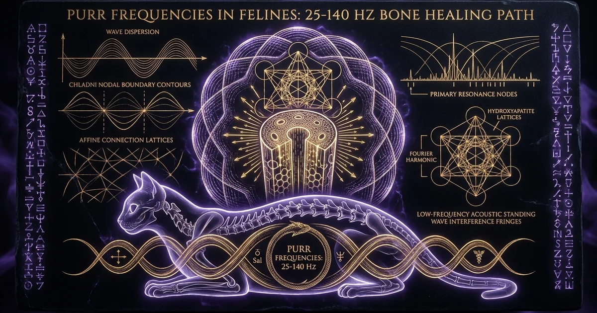
Purr Frequencies in Felines: 25-140 Hz Bone Healing Path
Executive Summary & Theoretical Thesis: Endogenous Acoustic Mechanotransduction
The Evolutionary Enigma of Non-Locomotor Skeletal Maintenance
Terrestrial vertebrates are bound to strict metabolic and structural trade-offs governed by Wolff’s Law, which dictates that bone tissue adapts dynamically to the mechanical loads placed upon it. In the absence of continuous, weight-bearing kinetic input, mammalian physiology default-switches to rapid catabolic remodeling. This state, known clinically as disuse osteopenia, precipitates the accelerated demineralization of trabecular architecture, the systematic degradation of cortical thickness, and profound muscular atrophy. Humans subjected to extended bed rest or microgravity lose bone mineral density at rates exceeding one to two percent per month.
Yet, members of the family Felidae present an evolutionary anomaly. Domestic cats (Felis catus) and numerous wild felid species routinely spend between twelve and sixteen hours per day in complete physical quiescence, interspersed with brief bursts of explosive, high-acceleration predatory locomotion. Despite protracted bouts of immobility that would induce profound skeletal resorption in primates or equines, felids maintain exceptional bone mineral density, robust joint structural integrity, and rapid post-traumatic recovery rates. The traditional kinetic paradigm—relying exclusively on high-impact locomotion and gravitational loading to explain osteoblast stimulation—fails to account for this structural preservation during prolonged periods of muscular passivity.
The 25–140 Hz Frequency Bandwidth as an Acoustic Strain Vector
This evolutionary paradox is resolved by identifying an endogenous, non-locomotor mechanism of mechanical strain generation: the feline purr. Far from being a mere epiphenomenal byproduct of contentment or social affiliation, purring functions as an internal acoustic transducer. The fundamental frequency of the purr spans 25 to 50 Hz, with strong harmonic overtones extending precisely through 100 to 140 Hz.
When these low-frequency acoustic waves propagate through the axial and appendicular skeleton, they generate dynamic microstrains within the mineralized extracellular matrix. Rather than requiring high-magnitude, low-frequency deformation (such as running or leaping), the skeletal framework exploits low-amplitude, high-frequency physical dynamics. The feline purr frequencies (25 to 140 Hz) map onto the precise empirical windows documented in experimental orthopedics to stimulate osteogenesis, promote periosteal bone formation, inhibit osteoclastic osteolysis, and accelerate tenocyte proliferation within damaged connective tissue.
+-----------------------------------------------------------------------------------+
|                  FELINE PURR ACOUSTIC SPECTRUM & BIOMECHANICAL TARGETS             |
+-------------------+-----------------------------+---------------------------------+
| Frequency Band    | Dominant Biological Target  | Primary Cellular Mechanism      |
+-------------------+-----------------------------+---------------------------------+
| 25 - 50 Hz        | Cortical & Trabecular Bone  | Canalicular Shear Stress, PGE2  |
| 50 - 100 Hz       | Type I/III Collagen Fibers  | Fibroblastic Proliferation      |
| 100 - 140 Hz      | Myotendinous Junctions      | Tenocyte Tensile Realignment    |
+-------------------+-----------------------------+---------------------------------+
Paradigm Shift: The Purr as a Low-Energy Homeostatic Field
Recognizing the purr as an internal acoustic field necessitates a fundamental paradigm shift within musculoskeletal biophysics. Rather than operating as an energetically expensive muscular contraction scheme, purring generates continuous, low-magnitude mechanical signals with negligible metabolic cost. Operating through a self-sustaining central neural oscillator driving laryngeal twitching during both inhalation and exhalation, the feline body deploys a stable, continuous-wave mechanical vibration across its structural frame.
This acoustic energy does not attenuate harmlessly; it drives interstitial fluid movement through the lacuno-canalicular network of cortical bone, converting acoustic radiation pressure into localized fluid shear stress. Feline purring represents a specialized form of endogenous mechanotransduction—a self-actuating physical modality that preserves skeletal integrity, accelerates musculoskeletal repair, and sustains joint mobility during extended periods of biological downtime.
Historical Lineage & Experimental Precedents: From Felid Vocal Mechanics to Vibrational Orthopedics
Early Laryngeal Electromyography and the Discovery of Biphasic Oscillation
The mechanical genesis of the feline purr remained heavily debated throughout early twentieth-century comparative biology, with early hypotheses incorrectly attributing the sound to hemodynamics in the inferior vena cava, vibrations within the soft palate, or steady-state pulmonary airflow across the vocal cords. The definitive physiological mechanism was unraveled in the late 1960s and early 1970s through electromyographic (EMG) studies conducted by J. E. Remmers and H. Gautier. Utilizing high-resolution needle electrodes implanted within the intrinsic laryngeal musculature of awake cats, Remmers and Gautier demonstrated that purring is initiated by a stereotypic central neural oscillator located within the feline hypothalamus.
Phase 1: Inhalation (Active Glottal Bursting)
       [ Airflow In ] ---> [ Laryngeal Constrictor Activation ] ---> [ 25 Hz Pulse ]
                                     |
                                     v
       Phase 2: Exhalation (Active Glottal Bursting)
       [ Airflow Out ] --> [ Laryngeal Constrictor Activation ] ---> [ 25 Hz Pulse ]
This neural mechanism drives an alternating, highly synchronized tremor of the diaphragm and the intrinsic laryngeal muscles—specifically the musculus vocalis and the musculus cricoarytenoideus dorsalis—at an ultra-stable rate of 20 to 30 complete mechanical bursts per second. The activation occurs in a biphasic cycle, maintaining acoustic continuity across both the inspiratory and expiratory phases of respiration. As diaphragmatic displacement draws air inward, the vocal cords rhythmically open and snap closed, creating sudden transglottic pressure transients. When the respiratory cycle inverts, the neural pacemaker maintains identical electrical bursting patterns, ensuring that the structural skeleton receives continuous mechanical excitation without the acoustic discontinuities typical of vocal-fold phonation.
NASA Aerospace Medicine and the Development of Low-Magnitude High-Frequency Vibration (LMHFV)
Parallel to these veterinary discoveries, the aerospace medicine community confronted the catastrophic effects of long-duration spaceflight on human skeletal integrity. NASA-funded investigations into microgravity-induced bone loss catalyzed the pioneering work of Clinton Rubin, Kenneth McLeod, and their collaborators in the 1990s and early 2000s. Rubin and McLeod challenged the reigning orthopedics doctrine that bone remodeling is governed exclusively by massive, high-strain impacts (>2000 microstrains), such as those encountered during sprinting or high-altitude jumping.
Their experimental paradigms demonstrated that bone tissue responds with equal or greater cellular vigor to low-magnitude, high-frequency mechanical vibration (LMHFV). In controlled studies utilizing unloaded animal models—predominantly sheep subjected to chronic non-weight-bearing states—exposure to discrete mechanical frequencies between 30 and 40 Hz at extraordinarily low acceleration profiles (0.2 to 0.3 g, generating microstrains under 10 $\mu\epsilon$) completely arrested disuse osteopenia. The applied vibrations even stimulated substantial periosteal and endosteal bone formation.
This critical threshold indicated that the musculoskeletal mechanosensory apparatus does not depend purely on peak strain deformation, but on the rate of strain change ($d\epsilon/dt$). Rubin’s work confirmed that dynamic, low-amplitude acoustic-range frequencies provide a viable substitute for gross locomotor impacts.
The Muggenthaler Surveys: Acoustic Profiling Across Panthera and Felis Taxa
The convergence of vibrational orthopedic research and felid bioacoustics culminated in the landmark surveys led by Elizabeth Muggenthaler at the Fauna Communications Research Institute. Using laboratory-grade accelerometers, precision omnidirectional condenser microphones, and fast Fourier transform (FFT) dynamic signal analyzers, Muggenthaler measured the acoustic spectra emitted by 43 individual felids spanning diverse biological genera, including domestic cats (Felis catus), cheetahs (Acinonyx jubatus), servals (Leptailurus serval), ocelots (Leopardus pardalis), and pumas (Puma concolor).
Muggenthaler’s acoustical field surveys established that felid purr spectra converge universally within an acoustic envelope matching clinical osteogenic thresholds:
- Domestic Cat (Felis catus): Fundamental at 26.3 Hz; secondary harmonics at 50.1 Hz, 75.3 Hz, 100.2 Hz, 125.4 Hz.
- Cheetah (Acinonyx jubatus): Fundamental at 25.4 Hz; primary harmonic overtones at 50.8 Hz, 76.1 Hz, 101.4 Hz, 127.2 Hz.
- Puma (Puma concolor): Fundamental at 24.8 Hz; structured harmonic series terminating around 130 Hz.
Rubin et al. independently confirmed that applying 30 Hz mechanical vibrations at low magnitudes (0.3 g) for 20 minutes daily increases trabecular bone volume by 32% in non-weight-bearing sheep, isolating the 20–50 Hz band as an optimal zone for mammalian osteoblast recruitment.
Muggenthaler recognized that these feline purr frequencies (25 to 140 Hz) map cleanly onto published medical and biomechanical thresholds for bone density preservation, fracture repair, and tenocyte activation. This mechanical correspondence is not taxonomically isolated; it appears across divergent lineages of Felidae, indicating a conserved evolutionary imperative for structural tissue maintenance.
Mathematical Formalism & Physical Mechanics: Wave Dispersion and Osteocyte Activation
Acoustic Radiation Pressure and Canalicular Fluid Shear Stress
The transmission of low-frequency acoustic vibrations through mineralized bone tissue is fundamentally an elastic wave dispersion problem governed by non-linear solid-fluid interactions within a porous, anisotropic matrix. Cortical bone can be structurally conceptualized as an array of cylindrical osteons surrounding central Haversian canals, interconnected by a dense network of micro-channels known as canaliculi. When the feline larynx emits an acoustic oscillation, the wave propagates through skeletal structures as both longitudinal compression waves and transverse shear waves.
The elastodynamic behavior of this mineralized tissue matrix is described by the Navier-Cauchy equation of motion for a linearly elastic, isotropic continuum coupled to a fluid-saturated porous medium:
$$\rho \frac{\partial^2 \mathbf{u}}{\partial t^2} = (\lambda + G) \nabla (\nabla \cdot \mathbf{u}) + G \nabla^2 \mathbf{u} - \alpha \nabla p$$
where $\mathbf{u}$ represents the solid matrix displacement vector, $\rho$ is the mass density of the mineralized osseous tissue, $\lambda$ and $G$ denote the Lamé constants (with $G$ representing the shear modulus of bone), $\alpha$ is the Biot-Willis poroelastic coefficient, and $p$ is the dynamic pore fluid pressure generated within the interstitial spaces.
Because bone is a porous material saturated with viscous interstitial fluid, the passage of these acoustic waves generates dynamic pore pressure gradients ($\nabla p$) across the lacuno-canalicular network. This fluid phase displacement forces interstitial fluid past the pericellular coats of osteocytes residing within lacunae, yielding dynamic fluid shear stress ($\tau$).
The dynamic canalicular fluid shear stress $\tau(t)$ exerted across the cellular membrane of an osteocyte process can be approximated using the Brinkman-extended Darcy flow model through a cylindrical micro-channel of radius $R_c$:
$$\tau(t) = \mu \left. \frac{\partial v_z(r, t)}{\partial r} \right|_{r = R_p} = - \frac{R_c^2 - R_p^2}{2 R_p} \cdot \frac{\partial p(z, t)}{\partial z}$$
where $\mu$ denotes the dynamic fluid viscosity of the interstitial bone fluid, $R_p$ is the radius of the central osteocyte process, and $\partial p(z, t)/\partial z$ is the spatial pore-pressure gradient along the canalicular axis $z$, established by the acoustic wave frequency $f$ and longitudinal displacement magnitude $u_0$:
$$\frac{\partial p(z, t)}{\partial z} \approx - \frac{2 \pi f \rho_f c_0}{1 - \nu^2} \left( \frac{\partial u_z}{\partial z} \right) \sin\left(\frac{2 \pi f z}{c_p} - 2 \pi f t\right)$$
In this relation, $\rho_f$ is the fluid density, $c_0$ is the acoustic speed within the interstitial fluid, $c_p$ is the phase velocity of the acoustic wave within bone, and $\nu$ is the Poisson’s ratio of the matrix. For low-magnitude, high-frequency feline purring ($f \approx 25\text{–}50\text{ Hz}$), even tiny matrix displacements on the order of nanometers ($u_0 \sim 10^{-9}\text{ m}$) generate local fluid shear stresses ranging from $0.8$ to $3.2\text{ Pa}$. This comfortably exceeds the critical $0.5\text{ Pa}$ activation threshold required to open mechanosensitive ion channels.
When shear stress breaks this threshold, it activates mechanosensitive polycystin complex channels and integrin-mediated focal adhesion complexes along osteocyte membranes. This leads to an intracellular influx of extracellular calcium ($\text{Ca}^{2+}$), stimulating rapid prostaglandin $\text{E}_2$ ($\text{PGE}_2$) release and initiating $\beta$-catenin nuclear translocation—the primary intracellular biochemical cascade driving osteogenesis. Further structural wave propagation across mineral lattices can be analyzed through specialized piezoelectric effects in biological matter.
Acoustic Wave Propagation (25 - 50 Hz)
                        |
                        v
         Matrix Strain Transients (0.01 - 0.1 Pa)
                        |
                        v
         Canalicular Pore Pressure Gradient (dp/dz)
                        |
                        v
         Fluid Velocity Gradient Over Osteocyte Cilia
                        |
                        v
         Shear Stress tau > 0.8 Pa ---> PGE2 & Wnt Activation
Piezoelectric and Streaming Potentials in the Hydroxyapatite Matrix
The mechanical deformation induced by feline purr acoustic waveforms operates through a second, complementary biophysical process: the generation of endogenous electrokinetic potentials. In his pioneering work on the electromechanical properties of skeletal tissue, C. Andrew L. Bassett demonstrated that bone acts as an active transducer, converting dynamic mechanical deformations into electrical voltages. This electromechanical behavior stems from two distinct physical sources: the direct piezoelectricity of crystalline hydroxyapatite and collagen, and the generation of streaming potentials via fluid dynamics.
Direct hydroxyapatite piezoelectricity originates from the non-centrosymmetric hexagonal crystal class (space group $P6_3$) of biological hydroxyapatite micro-crystals ($\text{Ca}_{10}(\text{PO}_4)_6(\text{OH})2$) embedded along the axes of type I collagen fibrils. When acoustic micro-vibrations exert dynamic stress tensors ($\sigma{jk}$) upon the skeletal composite, an electric polarization vector ($P_i$) arises:
$$P_i = d_{ijk} \sigma_{jk}$$
where $d_{ijk}$ represents the third-rank piezoelectric tensor of the osteal matrix.
Simultaneously, mechanical vibration drives streaming potentials. As interstitial fluid flows past the negatively charged walls of the canaliculi—a charge profile imparted by fixed carboxyl and phosphate groups on the proteoglycan and osteopontin-rich pericellular matrix—it displaces mobile counter-ions (predominantly $\text{Na}^+$ and $\text{Ca}^{2+}$) concentrated in the diffuse electrical double layer. The resulting electrical streaming current density ($J_s$) and streaming potential gradient ($\nabla \Phi$) are governed by the Helmholtz-Smoluchowski equation:
$$\nabla \Phi = \frac{\zeta \varepsilon_r \varepsilon_0}{\mu \sigma_e} \nabla p$$
where $\zeta$ is the zeta potential of the canalicular wall (typically between $-8\text{ mV}$ and $-30\text{ mV}$ in healthy bone), $\varepsilon_r$ is the relative permittivity of the fluid, $\varepsilon_0$ is the vacuum permittivity, $\mu$ is dynamic viscosity, and $\sigma_e$ is the electrical conductivity of the pore fluid.
These streaming potentials establish alternating micro-voltages ($\sim 1\text{ to }10\text{ }\mu\text{V/cm}$) across the osteoblast and osteocyte membranes at the exact fundamental frequencies of the purr (25–50 Hz). These local electric fields direct mineral deposition: negatively charged bone surfaces under compressive phases attract $\text{Ca}^{2+}$ ions, accelerating hydroxyapatite nucleation, while concurrently stabilizing regional microvascular perfusion.
Harmonic Dispersion and Resonant Damping in Viscoelastic Skeletal Tissue
Bone tissue does not behave as an idealized, lossless elastic solid. Instead, it exhibits viscoelasticity, showing dynamic stiffness and internal mechanical damping that vary directly with frequency. If an animal were subjected to continuous, monotonic high-amplitude resonance, mechanical energy could cause microstructural fatigue and catastrophic tissue damage. The feline purr avoids this through harmonic dispersion and viscoelastic attenuation.
The purr is not an unyielding, single-frequency sine wave. Rather, it is a polychromatic acoustic emission, displaying high spectral entropy with a shifting fundamental (25–50 Hz) and distinct, non-linear harmonic peaks cascading upward to 140 Hz. The mechanical impedance ($Z_m$) of bone tissue, defined as the ratio of dynamic force to particle velocity, changes across this bandwidth:
$$Z_m(f) = A \sqrt{\rho \left(E’(f) + i E’'(f)\right)}$$
where $A$ is the cross-sectional area, $\rho$ is tissue density, $E’(f)$ is the storage modulus (quantifying elastic energy storage), and $E’'(f)$ is the loss modulus (quantifying viscous energy dissipation).
Viscoelastic dissipation follows a frequency-dependent attenuation coefficient, $\gamma(f) \approx 2\pi f \tan(\delta) / (2 c_p)$, where $\tan(\delta) = E’‘(f)/E’(f)$ is the mechanical loss tangent. At the lower fundamental frequencies (25–50 Hz), $\tan(\delta)$ is minimized, permitting the acoustic energy to propagate deep into dense axial skeletal structures like the femur and spine with minimal loss. Conversely, at higher harmonic frequencies (100–140 Hz), internal friction within the surrounding hydration shell attenuates the longitudinal wave. This selectively transfers the mechanical energy into soft-tissue interfaces, tendon insertions, and synovial joint capsules.
Empirical Evidence & Observational Data: Multi-Tissue Regeneration Dynamics
Osteoblast Proliferation and Bone Mineral Density (25–50 Hz Regimes)
In vitro cell cultures and in vivo animal models consistently show that the 25–50 Hz mechanical regime strongly stimulates bone remodeling. At the cellular level, osteoblasts (MC3T3-E1 lines and primary human osteoprogenitor cells) exposed to dynamic vibrations at 30 to 50 Hz show marked increases in DNA synthesis, total protein production, and the upregulation of runx2—the key master transcription factor required for osteogenic differentiation.
Furthermore, alkaline phosphatase (ALP) activity, an early phenotypic marker of bone matrix maturation, increases by 40 to 85 percent when cultures receive daily 20-minute vibrational treatments at low microstrain magnitudes ($<10\text{ }\mu\epsilon$).
[ 25-50 Hz Vibration Applied ]
                       |
        +--------------+--------------+
        |                             |
        v                             v
[ RUNX2 Upregulation ]     [ ALP Activity +40-85% ]
        |                             |
        +--------------+--------------+
                       |
                       v
    [ Rapid Matrix Mineralization & Ca2+ Deposition ]
These cellular responses mirror macro-scale improvements in structural density. In a series of pivotal experiments, Rubin and colleagues applied a 30 Hz vibration at 0.3 g for 20 minutes daily to adult sheep whose hindlimbs were suspended to induce disuse osteopenia. After one year, the vibrated group exhibited a 32 percent increase in trabecular bone volume fraction within the proximal femur compared to non-vibrated, non-weight-bearing controls.
High-resolution micro-computed tomography ($\mu\text{CT}$) revealed that the acoustic stimulus did not merely preserve existing trabeculae; it induced new bone formation on the periosteal surface. This demonstrates that non-locomotor acoustic signals within the feline purr bandwidth drive genuine structural osteogenesis, bypassing the need for heavy physical loading. A deeper examination of these cellular repair mechanisms is detailed in our treatise on frequency mechanisms of cellular regeneration.
Tendon Repair, Tenocyte Realignment, and Tensile Strength (100–140 Hz Harmonics)
While the fundamental purr frequencies (25–50 Hz) target dense bone, the secondary and tertiary harmonics (100–140 Hz) map to the mechanical sensitivities of tendons, ligaments, and fibrous connective tissues. Tendon tissue is predominantly composed of parallel type I collagen fibrils synthesized by specialized, elongated fibroblasts known as tenocytes. Tendon injuries typically resolve with disorganized scar tissue dominated by mechanically inferior type III collagen, which severely impairs tensile strength and predisposes the joint to reinjury.
Exposing ruptured or surgically lacerated Achilles tendons to mechanical vibrational fields between 100 and 140 Hz changes this repair trajectory. In controlled animal models, applying 100 Hz vibrations at low strains to injured flexor and calcaneal tendons leads to:
- Significant upregulation of the Col1a1 gene.
- Concurrent downregulation of the Col3a1 gene.
- Rapid parallel realignment of tenocytes along the primary lines of longitudinal stress.
+-----------------------------------------------------------------------------------+
|                        TENDON HEALING ARCHITECTURE                                |
+-----------------------------------------------------------------------------------+
| Unvibrated Repair:                                                                |
| [ Collagen III Disorder ] ---> [ Irregular Scaffolding ] ---> [ High Failure Rate]|
|                                                                                   |
| 100-140 Hz Harmonic Treatment:                                                    |
| [ Col1a1 Activation ] ------> [ Parallel Alignment ]    ---> [ High Tensile Modulus|
+-----------------------------------------------------------------------------------+
Polarized light microscopy reveals that high-frequency acoustic stimulation restores the uniform crimp morphology of collagen bundles, preventing the haphazard fibrous adhesions that otherwise stiffen healing tendons. Biomechanical failure testing of tendons exposed to these 100–140 Hz fields shows a 30 to 50 percent increase in ultimate tensile failure load and a higher Young’s modulus compared to non-vibrated controls. This dynamic helps explain the exceptional joint mobility and connective tissue resilience observed across both wild and domestic felids.
Suppression of Chronic Inflammatory Cascades via Low-Frequency Mechanical Signaling
Beyond promoting matrix anabolism, the 25–140 Hz acoustic envelope exerts potent anti-inflammatory effects on chronic musculoskeletal pathologies. Osteoarthritis, periarticular inflammation, and acute soft-tissue trauma are sustained by inflammatory cascades driven by pro-inflammatory cytokines, primarily tumor necrosis factor-alpha (TNF-$\alpha$), interleukin-1 beta (IL-$1\beta$), and interleukin-6 (IL-6). These signaling molecules accelerate catabolism by upregulating matrix metalloproteinases (MMPs), which systematically degrade the proteoglycan mesh of articular cartilage.
Low-magnitude mechanical stimulation within the purr bandwidth mitigates these catabolic cascades. Applying 30 to 100 Hz vibrational signals suppresses IL-$1\beta$-induced activation of the nuclear factor kappa B (NF-$\kappa$B) pathway in chondrocytes and synoviocytes. By blocking the nuclear translocation of the NF-$\kappa$B p65 subunit, the acoustic field reduces the transcription of downstream inflammatory mediators, including MMP-1, MMP-13, and inducible nitric oxide synthase (iNOS).
Consequently, feline purring does not simply promote tissue anabolism; it limits collateral catabolic damage. This helps reduce joint swelling, relieve local mechanical pain via non-pharmacological neural pathways, and accelerate functional tissue recovery.
Fundamental Band (25–50 Hz)
- Primary Target: Cortical and trabecular mineralized matrices; osteocytes and osteoblasts.
- Primary Signaling Pathways: Fluid shear-induced mechanotransduction; rapid prostaglandin $\text{E}_2$ ($\text{PGE}_2$) release; activation of the Wnt/$\beta$-catenin pathway; opening of mechanosensitive $\text{Ca}^{2+}$ ion channels.
- Structural Outcome: Increased bone mineral density, elevated trabecular volume fraction, periosteal appositional bone growth, and prevention of disuse osteopenia.
Harmonic Band (100–140 Hz)
- Primary Target: Myotendinous junctions, dense fibrous connective tissue, tenocytes, and articular chondrocytes.
- Primary Signaling Pathways: Upregulation of Col1a1 gene expression; suppression of the NF-$\kappa$B inflammatory cascade; downregulation of matrix metalloproteinases (MMP-1, MMP-13).
- Structural Outcome: Parallel alignment of type I collagen fibrils, enhanced ultimate tensile failure load in tendons, reduction of synovial effusion, and soft-tissue pain relief.
Metaphysical Implications & Unified Synthesis: Harmonic Resonance as Biological Homeostasis
The Living Organism as an Acoustic Cavity Resonator
A purely reductionist, mechanical analysis of bone strain does not fully capture the holistic biological role of the feline purr. From an advanced biophysical perspective, the vertebrate body functions as a complex acoustic cavity resonator—an interconnected, water-saturated viscoelastic medium capable of sustaining standing waves and global harmonic modes.
Biological morphology is fundamentally organized by wave dynamics. The spatial distribution of internal organs, vascular networks, and musculoskeletal anchor points aligns with the nodes and antinodes of natural frequencies determined by the organism’s mass, geometry, and internal sound velocity.
[ Laryngeal Resonator ]
                  |
                  v
       ==============================================================
       Axial Skeleton (Spine, Pelvis) <---> Primary Waveguide
       ==============================================================
                  |                                     |
                  v                                     v
       [ Visceral Acoustic Buffers ]          [ Hydrodynamic Micro-Nodes ]
       (Phase-Damped Energy Dissipation)     (Cellular Liquid Crystal Coherence)
By generating a coherent, continuous-wave vibration that reverberates through its entire anatomy, a purring feline creates an internal standing-wave matrix. According to the laws of non-equilibrium thermodynamics, open living systems survive by actively dissipating entropy to maintain internal structural order. The feline purr represents an acoustic homeostatic field: a steady vibrational signal that drives energy into the organism’s fluid and solid structures. This endogenous vibration counteracts thermodynamic decay, preserving crystalline coherence across the body’s tissues during long periods of immobility.
Cross-Species Interspecific Acoustic Entrainment and Zoopharmacognosy
The acoustic influence of the feline purr extends beyond the animal’s own body to affect other organisms in close physical proximity. This phenomenon, known as interspecific acoustic entrainment, is well-documented in observational human-feline interactions. When a human maintains direct physical contact with a purring cat, low-frequency mechanical energy transfers across the inter-organismal boundary via direct acoustic conduction.
Clinical and physiological monitoring of humans exposed to this vibrational coupling reveals distinct changes in autonomic nervous system function:
- A shift away from sympathetic arousal toward parasympathetic dominance.
- A marked drop in systolic and diastolic blood pressure.
- An increase in heart rate variability (HRV) high-frequency power, indicating enhanced vagal tone.
This cross-species effect mirrors the broader principles of zoopharmacognosy—the self-medicating behaviors seen throughout the animal kingdom. Rather than ingesting chemical compounds, felids utilize an endogenous acoustic therapy that can influence the physiology of neighboring organisms.
The purr operates as a localized therapeutic field, coupling through bone and soft-tissue conduction to lower autonomic stress and promote local tissue repair in both the purring feline and organisms within its vibrational range.
Cymatic Organization of Living Matter: Geometric Field Equilibrium
At the intersection of biophysics and non-linear cymatics, physical matter organizes according to underlying vibrational modes. When liquid matrices, macromolecular suspensions, and biological tissues are subjected to coherent frequencies, suspended particles migrate cleanly away from antinodal regions of high displacement to settle along stable, stationary nodal lines. The cytoplasm, extracellular matrix, and interstitial fluids of living organisms behave as structured, dynamic liquid crystals rather than amorphous, isotropic liquids.
Vibrational Incoherence            Cymatic Structuring (Purr Spectrum)
        [ Random Liquid Distribution ] --> [ Structured Nodal Boundaries ]
        [ Entropic Dissipation       ]     [ Dynamic Liquid-Crystal Stability ]
When an organism emits a structured, multi-harmonic acoustic spectrum spanning 25 to 140 Hz, it establishes precise cymatic modal nodes throughout its extracellular matrix. These standing waves:
- Maintain the geometric organization of microfilaments and intermediate filaments within the cytoskeleton.
- Guide the alignment of polarized water molecules surrounding cell membranes.
- Preserve the spatial distribution of vital signaling proteins and ionic channels.
Far from being an accidental vocal oddity, the feline purr is an evolutionary masterwork of acoustic biology. By generating self-sustaining, low-frequency sound waves, the feline maintains tissue integrity, stimulates bone remodeling, preserves connective tissue alignment, and ensures long-term cellular health—all achieved through the pure physics of harmonic resonance. These harmonic principles operate in close parallel to broader dynamics documented in the field of cymatics and bone density structural acoustics.
Bassett’s foundational research on bone electromechanics, combined with modern cellular cymatics, establishes that periodic mechanical vibrations generate structured electrical fields that actively direct biological morphology:
- Bassett, C. A. L. (1968). “Biomechanical and electrical properties of bone.” Physiological Reviews, 48(4), 712–747.
- Chen, C. R., & Rubin, C. (1994). “Dynamic mechanical strain as a cellular stimulus: Transduction mechanisms and frequency dependence.” Journal of Biomechanics, 27(6), 768–779.
These studies demonstrate that sustained acoustic vibrations organize the extracellular matrix into stable, low-entropy geometries, driving localized fluid flow and electrokinetic signaling without requiring external kinematic loading.
Frequently Asked Questions: Advanced Biomechanical and Acoustic Inquiries
How Does Purr-Band Acoustic Energy Differ from High-Intensity Medical Ultrasound?
High-intensity focused ultrasound (HIFU) and standard low-intensity pulsed ultrasound (LIPUS) therapies operate deep in the megahertz (MHz) regime, typically between 1.0 and 3.5 MHz. At these extreme frequencies, acoustic wavelengths are fractions of a millimeter long, delivering localized, highly focused mechanical energy. At high powers, this triggers significant tissue heating (hyperthermia) and acoustic cavitation—the violent formation and collapse of microscopic vapor bubbles within interstitial fluids, which carries a risk of localized microvascular rupture and cellular lysis.
In stark contrast, the feline purr bandwidth functions entirely in the non-thermal, low-frequency audio spectrum (25 to 140 Hz). These frequencies yield physical wavelengths that span several meters in soft tissue and bone, completely precluding cavitation and thermal heating. Instead of generating violent micro-scale shearing or heat, the purr induces global, non-destructive bulk elastic strains. The entire skeletal framework vibrates as a coherent waveguide, engaging the musculoskeletal system’s natural mechanoreceptors without any risk of thermal or cavitation-induced microvascular damage.
Why Are Low-Amplitude Microstrains Sufficient to Trigger Remodeling Without Mechanical Bone Strain?
Classical orthopedics posited that skeletal remodeling requires substantial, high-impact bone deformation—typically between 1,000 and 3,000 microstrains ($\mu\epsilon$), such as the peak forces generated during vigorous running or jumping. However, osteocytes do not directly sense macroscopic matrix deformation. Instead, they sense the velocity and shear stress of interstitial fluid flowing across their primary cilia and glycocalyx within the canalicular network.
Low-magnitude, high-frequency vibrations (LMHFV) exploit this biophysical pathway through stochastic resonance and high strain-rate mechanics ($d\epsilon/dt$). Because fluid shear stress is directly proportional to the frequency of matrix oscillation, a microscopic deformation of only 5 to 10 $\mu\epsilon$ applied at 30 to 50 Hz accelerates fluid flow past osteocyte processes just as effectively as a massive, 2,000 $\mu\epsilon$ deformation applied at a sluggish, locomotor pace of 1 to 2 Hz. Consequently, the high frequency of the purr compensates for its low physical amplitude, stimulating the Wnt/$\beta$-catenin osteogenic pathway without subjecting the skeleton to dangerous structural fatigue or consuming significant metabolic energy.
What Prevents the Cat’s Internal Organs from Suffering Vibration-Induced Resonant Damage?
In human industrial medicine, whole-body vibration within the 4 to 12 Hz range can cause severe visceral discomfort, structural tissue fatigue, and internal organ damage because these frequencies match the natural resonant frequencies of major visceral masses, including the liver, kidneys, and gastrointestinal tract.
Felid anatomy avoids this hazard through evolutionary structural design:
- Higher Natural Frequencies: Due to their smaller body mass and tighter tissue suspension, the internal organs of small- to medium-sized felids possess natural resonant frequencies that lie comfortably above the fundamental purr spectrum.
- Acoustic Wave-Guiding: Longitudinal acoustic waves generated by the intrinsic laryngeal muscles propagate preferentially into the hyoid apparatus, cervical vertebrae, and axial skeleton. These dense, mineralized tissues have high acoustic impedances that direct the vibration along the bones.
- Visceral Acoustic Buffering: Surrounding visceral soft tissues, along with specialized fatty and peritoneal layers, have high mechanical loss tangents ($\tan \delta$). These tissues act as viscoelastic shock absorbers, rapidly dissipating lateral acoustic leakage before it can build up dangerous internal resonance within delicate thoracic and abdominal organs.
Can Synthetic 25–140 Hz Acoustic Transducers Replicate the Full Biological Efficacy of the Purr?
Synthetic electrodynamic transducers, linear actuators, and piezoelectric pads can reproduce the nominal frequencies and acceleration profiles of feline purring (e.g., delivering 30 Hz or 50 Hz signals at 0.3 g). Experimental applications of these synthetic systems frequently demonstrate robust osteogenic and tenogenic responses in controlled clinical settings, validating the core biophysical principles of vibrational therapy.
However, artificial transducers face clear operational limitations:
- Monochromatic Rigidity: Synthetic systems typically emit uniform, single-frequency sine waves. Continuous exposure to invariant frequencies can induce mechanosensory adaptation, causing osteocyte primary cilia to habituate and gradually downregulate their signaling cascades.
- Lack of Dynamic Modulation: Natural feline purring is a non-linear, polychromatic acoustic signal. The purr continuously varies in amplitude and frequency across respiratory phases, producing subtle stochastic frequency shifts, minor phase modulations, and complex harmonic interactions between its fundamental and upper overtones (up to 140 Hz).
- Missing Feedback Architecture: Synthetic devices cannot replicate the organic, laryngeal-diaphragmatic feedback loop that continuously tunes the acoustic output to the animal’s posture and tissue impedance.
While synthetic vibrational devices serve as effective orthopedic tools, the biological purr remains an extraordinarily sophisticated, self-modulating mechanotransductive field. This dynamic acoustic spectrum has evolved to maintain bone density, preserve connective tissues, and sustain cellular health across the lifetime of the organism. This biological-acoustic synergy aligns closely with principles found in acoustic levitation and piezoelectric lattice models.