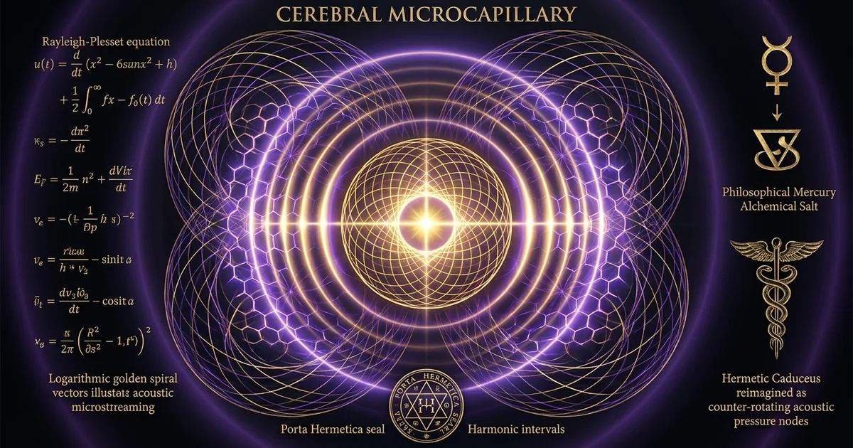
Sonic Cavitation in Targeted Drug Delivery Across the BBB
Executive Summary & Theoretical Thesis
The Neurovascular Interface and the Macromolecular Transport Dilemma
The vertebrate central nervous system is sequestered behind the blood-brain barrier (BBB), a continuous, non-fenestrated brain capillary endothelial lining characterized by high-density inter-endothelial junctional complexes. These microvascular networks generate a baseline transendothelial electrical resistance (TEER) exceeding 1500 to 2000 $\Omega \cdot \text{cm}^2$. This physiological insulation eliminates passive solute flux, effectively excluding more than 98% of small-molecule neuropharmaceuticals and 100% of high-molecular-weight therapeutics—including monoclonal antibodies, recombinant proteins, viral vectors, and liposomal carriers—from entering the cerebral parenchyma.
The maintenance of this selective semipermeable state is regulated by the cooperative architecture of the neurovascular unit (NVU). Brain microvascular endothelial cells (BMECs) rest upon a continuous basal lamina, enveloped by pericytes and the terminal foot processes of protoplasmic astrocytes. The physical impermeability of this interface is governed by specialized multiprotein assemblies comprising transmembrane tight junction (TJ) proteins—chiefly claudin-5 and occludin—anchored to the actin cytoskeleton via the peripheral membrane adaptor protein zonula occludens-1 (ZO-1).
Efforts to bypass this protective barrier in neuro-oncology have historically relied on hyperosmolar disruptions, such as the intra-arterial administration of hypertonic mannitol, or invasive stereotactic interventions. Hyperosmolar regimens induce widespread, non-selective endothelial cell shrinkage, precipitating global paracellular opening accompanied by broad influx of neurotoxic plasma proteins, transient seizures, and diffuse vasogenic edema. Consequently, targeted drug delivery across this bio-interface requires a non-invasive, spatially restricted, and transiently titratable physical mechanism capable of disengaging junctional complexes without destroying the underlying vascular or parenchymal architecture.
Acoustic Cavitation Regimes: Stable Oscillation versus Inertial Collapse
The strategic deployment of transcranial focused ultrasound (FUS) combined with intravenously administered pre-formed gas-filled microbubbles provides a targeted, non-thermal biomechanical solution to the macromolecular transport dilemma. When exposed to an alternating acoustic pressure field characterized by longitudinal waves traversing the brain parenchyma, gas-filled microbubbles act as local acoustic resonators, an effect known as acoustic cavitation targeted oncology. The operational regime of these microbubbles is determined by the local peak negative acoustic pressure ($P_{\text{neg}}$) and the fundamental insonation frequency ($f$), codified nondimensionally through the mechanical index (MI).
Within the lower acoustic parameter spectrum, typically characterized by an MI ranging between 0.4 and 0.8, microbubbles undergo stable (non-inertial) cavitation. In this sustained regime, the microbubble shell sustains linear and non-linear volumetric oscillations about an equilibrium radius over hundreds of acoustic cycles without undergoing structural fragmentation. These volumetric expansions and contractions produce localized acoustic microstreaming fields in the adjacent plasma and exert secondary Bjerknes forces against the endothelial luminal membrane.
Conversely, when acoustic pressures exceed the threshold for inertial cavitation (typically $\text{MI} > 0.8 - 1.0$, depending on local vascular architecture and gas core mechanics), the inward inertia of the surrounding fluid dominates the bubble dynamics. The microbubble undergoes unbounded expansion followed by violent, asymmetric collapse, generating localized shockwaves, transient hyperthermal zones ($>5000\text{ K}$ at the collapse core), and high-velocity liquid microjets ($>100\text{ m/s}$). Within brain capillaries, where the lumen diameter ($5 - 8\text{ }\mu\text{m}$) approximates or is smaller than the resonant bubble excursion envelope, inertial cavitation induces mechanical rupture of the capillary wall, erythrocyte extravasation, microhemorrhage, and petechial parenchymal necrosis.
Targeted focused ultrasound microbubbles blood brain barrier drug delivery relies on maintaining strictly controlled stable cavitation fields. This approach operates below the inertial collapse threshold to harness continuous, non-destructive mechanical work within the microcapillary compartment.
Biophysical Trajectory: From Capillary Distension to Tight Junction Cleavage
The biophysical cascade that bridges acoustic insonation with molecular transport begins within the capillary lumen. As microbubbles undergo sustained volumetric oscillations, the boundary conditions imposed by the rigid or viscoelastic endothelial tube restrict spherical expansion. This geometric constraint redirects volumetric displacement into lateral and circumferential distension of the capillary wall, generating transient hoop stresses and periodic strain fields across the endothelial monolayer.
Concurrently, the steep velocity gradients established at the bubble-liquid boundary drive secondary acoustic microstreaming flows, producing steady fluid shear stresses ($\tau_w$) on the order of 10 to 100 Pa directly upon the endothelial glycocalyx and luminal plasma membrane. This mechanical loading activates local mechanoreceptors, triggering intracellular calcium ($\text{Ca}^{2+}$) influx, downstream activation of endothelial nitric oxide synthase (eNOS), and protein kinase C (PKC) signaling cascades.
The structural consequence of this mechanical transduction is the rapid, transient tight junction opening governed by the phosphorylation, spatial redistribution, and clathrin-mediated endocytosis of claudin-5 and occludin. Simultaneously, the adapter protein ZO-1 dissociates from its submembranous anchor sites, severing the mechanical link between the transmembrane seals and the actin-myosin cytoskeleton. This disengagement produces nanoscale paracellular apertures, allowing non-permeating systemic therapeutics to passively extravasate along their electrochemical and concentration gradients into the interstitium. The process preserves the integrity of the basal lamina and underlying astrocytic end-feet, establishing a reversible opening window that reseals over a 4-to-6-hour period as the transmembrane proteins recycle to the cell boundary.
The mechanical index (MI) serves as the primary metric for estimating non-thermal bioeffects in non-linear acoustic fields: $$\text{MI} = \frac{P_{\text{neg}}}{\sqrt{f}}$$ where $P_{\text{neg}}$ is the peak negative acoustic pressure in megapascals (MPa) and $f$ is the transducer center frequency in megahertz (MHz). In transcranial applications across the human neurovasculature:
- $\text{MI} < 0.4$: Sub-therapeutic window; insufficient acoustic radiation force and microstreaming to trigger claudin-5 disengagement.
- $0.4 \le \text{MI} \le 0.8$: Optimal non-thermal therapeutic window; supports sustained stable cavitation, yielding transient tight junction opening without microvascular disruption.
- $\text{MI} > 0.8$: Pathological threshold; induces inertial cavitation, localized liquid microjets, vessel rupture, petechial hemorrhage, and irreversible blood-brain barrier disruption.
Historical Lineage & Experimental Precedents
The Evolution from Surgical Craniectomy to Transcranial Phased Arrays
The concept of utilizing high-frequency sound waves to modulate central nervous system structures originated in the mid-20th century. Early pioneers, including John and William Fry in the 1950s, demonstrated that high-intensity focused ultrasound (HIFU) could produce localized lesions within deep brain nuclei to manage neurodegenerative tremors. However, these foundational experiments required an invasive surgical craniectomy.
The human calvarium presents a significant acoustic obstacle: human cranial bone possesses an acoustic impedance ($Z \approx 5 - 8 \times 10^6\text{ Rayl}$) substantially higher than that of water or brain tissue ($Z \approx 1.5 \times 10^6\text{ Rayl}$), coupled with a longitudinal wave propagation speed ($c_b \approx 2500 - 3500\text{ m/s}$) nearly double that of the soft tissue parenchyma ($c \approx 1540\text{ m/s}$). This impedance mismatch, combined with variable calvarial thickness, structural diploë layers, and high acoustic attenuation ($\alpha \sim 10 - 20\text{ dB/cm}$ at 1 MHz), historically resulted in severe acoustic wave dephasing, focal point defocusing, and thermal absorption that could burn the scalp and periosteum.
Overcoming this skull barrier necessitated two technological developments: the emergence of multi-element hemispherical phased-array transducers and the integration of computed tomography (CT)-derived acoustic ray-tracing algorithms. By mapping the bone density variations of individual calvaria via high-resolution pre-operative CT scans, clinicians can assign real-time phase and amplitude correction offsets to over 1000 independent piezoelectric transducer elements arrayed across a hemispherical helmet.
By applying discrete phase shifts ($\Delta \theta_i$) to the longitudinal waves emitted by each element, the distorted acoustic wavefronts constructively recombine at a designated sub-millimeter stereotactic coordinate deep within the cerebrum. This innovation shifted therapeutic ultrasound from an invasive, ablation-limited modality to a non-invasive platform for transcranial energy delivery.
The Landmark 2001 Hynynen Breakthrough: Microbubble Amplification
Despite the precision of transcranial phased arrays, early applications focused almost exclusively on thermal tissue coagulation, using high acoustic intensities ($>1000\text{ W/cm}^2$) to ablate intracranial targets. In 2001, Kullervo Hynynen and colleagues altered this paradigm by decoupling the bio-acoustic mechanism from macroscopic thermal ablation.
Hynynen recognized that the direct mechanical interaction of acoustic waves with biological tissue requires high pressures to overcome baseline physical thresholds. However, introducing pre-formed, systemically circulated gas microbubbles—originally developed as echocardiographic contrast agents—dramatically changed these biophysical constraints.
These lipid- or protein-shelled microbubbles contain dense, low-solubility fluorinated gases (such as octafluoropropane or sulfur hexafluoride) with diameters between 1 and 4 $\mu\text{m}$. Because of their high volumetric compressibility relative to the surrounding incompressible plasma, they serve as localized focal amplifiers of acoustic energy. Upon insonation by low-frequency acoustic fields, these microbubbles expand and contract, concentrating the ambient acoustic field within their immediate microvascular vicinity.
Hynynen’s experimental data showed that systemic administration of microbubbles lowered the acoustic power required to achieve blood-brain barrier permeabilization by up to three orders of magnitude. For the first time, focused ultrasound disrupted tight junction integrity non-thermally without heating intervening skull bone or adjacent brain parenchyma. Dynamic contrast-enhanced magnetic resonance imaging (DCE-MRI) confirmed localized, reversible extravasation of gadolinium-based contrast agents, marking the development of focused ultrasound microbubbles blood brain barrier drug delivery as an active field of therapeutic investigation.
Primary Reference: Hynynen, K., McDannold, N., Vykhodtseva, N., & Jolesz, F. A. (2001). Noninvasive MR Imaging-guided Focal Opening of the Blood-Brain Barrier in Rabbits. Radiology, 220(3), 640-646.
- Insonation Center Frequency: 1.5 MHz (calibrated for high-precision trans-dural targeting).
- Burst Length: 10 milliseconds.
- Pulse Repetition Frequency (PRF): 1 Hz.
- Total Sonication Duration: 20 seconds.
- Acoustic Contrast Agent: Optison (perfluorocarbon-filled human serum albumin-shelled microbubbles) dosed at 0.05 to 0.1 mL/kg.
- Primary Finding: Non-thermal, non-destructive focal opening of the blood-brain barrier demonstrated via gadolinium-DTPA signal enhancement on T1-weighted MRI, showing no parenchymal architecture disruption or ischemic neuronal necrosis at derated peak negative pressures below 0.8 MPa.
Clinical Translation: Phase I/II Clinical Trials in Glioblastoma Multiforme
Following extensive preclinical validation across rodent and non-human primate models, clinical translation of this technology focused on neuro-oncological conditions, particularly glioblastoma multiforme (GBM). Glioblastoma features profound intratumoral genetic heterogeneity, aggressive microvascular infiltration, and an intact or partially functional blood-tumor barrier (BTB) within infiltrating, non-enhancing margins where invasive tumor stem cells reside. Systemic chemotherapeutic agents, such as doxorubicin, paclitaxel, and macromolecular monoclonal antibodies, frequently fail clinically due to sub-therapeutic parenchymal penetration in these infiltrative boundaries.
Human clinical trials (e.g., NCT02253212 and NCT03616860) have verified the safety, feasibility, and spatial accuracy of non-invasive, MR-guided focused ultrasound (MRgFUS) systems, such as the ExAblate Neuro platform, alongside surgically implanted ultrasound arrays like the SonoCloud system. Initial phase I/II clinical trials led by Mainprize, Lipsman, and colleagues demonstrated that transcranial FUS sonication in human GBM patients receiving standard-of-care adjuvant chemotherapies produced immediate, spatially conformal contrast enhancement in previously non-enhancing target zones.
Serial DCE-MRI protocols and volumetric pharmacokinetic assays confirmed local increases in the concentration of systemically administered chemotherapeutic and immunotherapeutic compounds. Post-treatment neurological and radiological assessments showed no symptomatic intracranial hemorrhages, ischemic infarctions, or sustained neurocognitive declines, moving focused ultrasound microbubbles blood brain barrier drug delivery from an experimental concept into clinical oncology.
Mathematical Formalism & Physical Mechanics
The Rayleigh-Plesset-Noltingk-Neppiras-Poritsky (RPNNP) Formulation
The fundamental dynamics of an unconstrained, spherical gas bubble suspended in an infinite incompressible Newtonian liquid subjected to a dynamic acoustic pressure field are governed by the classical Rayleigh-Plesset equation. However, to model an encapsulated microbubble within the viscous, micro-confined geometry of a brain capillary, this formulation must be extended. It must account for shell viscoelastic properties, liquid compressibility, dynamic viscosity, surface tension, and the mechanical resistance imposed by the vessel wall.
Building on the theoretical frameworks established by Church (1995) and refined by Marmottant et al. (2005), the radial motion $R(t)$ of an encapsulated microbubble exposed to a time-varying longitudinal acoustic driving pressure $P_a(t) = -P_A \sin(\omega t)$ is described by the modified RPNNP differential equation:
$$\rho_L \left( R \ddot{R} + \frac{3}{2}\dot{R}^2 \right) = \left[ P_0 + \frac{2\sigma(R_0)}{R_0} \right] \left( \frac{R_0}{R} \right)^{3\gamma} - \frac{2\sigma®}{R} - \frac{4\mu_L \dot{R}}{R} - \frac{4\kappa_s \dot{R}}{R^2} - 4\chi_s \left( \frac{1}{R_0} - \frac{1}{R} \right) - P_0 - P_a(t) - P_{\text{wall}}(R, \dot{R}, R_{\text{cap}})$$
In this mathematical representation:
- $\rho_L$ represents the density of the surrounding liquid medium (blood plasma, $\sim 1025\text{ kg/m}^3$).
- $R_0$ denotes the quiescent, initial equilibrium radius of the microbubble ($1.0 - 2.5\text{ }\mu\text{m}$).
- $\gamma$ is the polytropic gas exponent, which transitions from isothermal ($\gamma = 1$) to adiabatic conditions depending on the Péclet number of the core gas.
- $\sigma®$ is the dynamic, state-dependent surface tension of the encapsulating lipid monolayer.
- $\mu_L$ is the dynamic shear viscosity of blood plasma ($\sim 1.2 - 1.5\text{ mPa}\cdot\text{s}$).
- $\chi_s$ represents the shell elastic compression modulus (governing resistance to areal strain, typically $0.1 - 1.5\text{ N/m}$).
- $\kappa_s$ is the surface dilatational shell viscosity (providing intrinsic dynamic energy dissipation, typically $10^{-8} - 10^{-7}\text{ kg/s}$).
- $P_{\text{wall}}$ represents the hydrodynamic boundary condition constraint imposed by the close proximity of the microvessel wall of radius $R_{\text{cap}}$, which increases effective fluid inertia and shifts the microbubble’s resonance frequency $\omega_0$ downward.
Primary Reference: Church, C. C. (1995). A theoretical study of cavitation generated by an acoustic pulse. The Journal of the Acoustical Society of America, 97(3), 1510-1521. Theoretical Framework: Church expanded the foundational Noltingk-Neppiras-Poritsky models to explicitly account for the continuous viscoelastic shell encapsulating modern lipid- and protein-based contrast agents. By isolating the energy dissipation terms: $$\Phi_{\text{dissipation}} = \frac{4\mu_L \dot{R}}{R} + \frac{4\kappa_s \dot{R}}{R^2}$$ Church demonstrated that non-linear shell compliance prevents premature inertial rupture across diagnostic and therapeutic pressure thresholds, establishing the analytical basis for predicting the stable oscillation band that prevents thermal parenchymal ablation.
When driving acoustic pressures remain beneath the fragmentation limit, this equation yields an oscillatory radius solution exhibiting fundamental harmonic components ($\omega$), sub-harmonic emissions ($\frac{1}{2}\omega$), and higher-order super-harmonics ($2\omega, 3\omega, \dots$). These non-linear oscillations generate targeted mechanical forces directly against the vascular endothelium.
Microvascular Fluid Mechanics: Acoustic Microstreaming and Shear Stress
Stable, non-linear microbubble radial oscillations within a fluid medium generate localized momentum transfers. These dynamics arise from the Reynolds stress tensor within the oscillating boundary layer surrounding the bubble interface. This secondary, steady circulatory fluid motion, known as acoustic microstreaming, induces steep velocity gradients near the cellular membrane.
The acoustic microstreaming velocity field $\mathbf{u}_s$ can be calculated through the time-averaged, non-linear Navier-Stokes equations governing an incompressible fluid. By separating the velocity into a first-order oscillatory component $\mathbf{u}_1$ and a second-order time-averaged steady streaming component $\mathbf{u}_2$:
$$\rho_L \langle (\mathbf{u}_1 \cdot \nabla) \mathbf{u}_1 \rangle = -\nabla \langle p_2 \rangle + \mu_L \nabla^2 \mathbf{u}_2$$
The resulting steady shear stress tensor acting on the endothelial cell surface located at distance $h$ from the oscillating bubble center is defined by:
$$\tau_w = \mu_L \left. \left( \frac{\partial u_{2,x}}{\partial y} \right) \right|_{y=0}$$
Analytical and numerical calculations show that for a microbubble oscillating stably at 1 MHz with a radial excursion amplitude $\Delta R/R_0 \approx 0.2 - 0.5$ within a microcapillary, local wall shear stresses ($\tau_w$) reach values between 10 and 100 Pa. These values exceed baseline physiological laminar fluid shear stresses within the cerebral microvasculature (typically $1 - 2\text{ Pa}$) by one to two orders of magnitude.
Simultaneously, the acoustic radiation force—derived from the primary longitudinal acoustic field interacting with the microbubble’s acoustic cross-section—pushes the bubble against the vessel wall:
$$\mathbf{F}{\text{ARF}} = -\frac{1}{2} \text{Re} \left{ \int{S} \langle p_1 \mathbf{n} \rangle , dS \right} = -\frac{2\pi P_A^2 R_0}{\rho_L \omega} \frac{\delta_{\text{tot}}}{(\omega_0^2/\omega - \omega)^2 + \delta_{\text{tot}}^2}$$
This acoustic-radiation-force presses the oscillating microbubble directly into the endothelial glycocalyx. This contact enhances the hydrodynamic shear stresses acting on local cell-cell junctions and transfers momentum directly to the plasma membrane.
Tight Junction Dissociation Kinetics and Paracellular Pore Hydrodynamics
The application of localized shear stress $\tau_w$ and circumferential strain $\epsilon_{\text{circ}}$ initiates the disengagement of the multiprotein complexes that form the tight junction seal. Claudin-5 molecules, which form the primary barrier to small molecules via homophilic and heterophilic interactions across the paracellular space, are anchored to the submembranous cytoskeleton via the PDZ-domain of ZO-1.
Upon receipt of the biomechanical stimulus, BMECs initiate a rapid phosphorylation cascade. Elevated intracellular shear triggers the phosphorylation of the regulatory light chain of myosin II and induces tyrosine phosphorylation of occludin (at residues Tyr398 and Tyr402) and claudin-5.
This biochemical modification breaks the electrostatic and conformational bonds linking the claudin-5 cytoplasmic tail to the ZO-1 scaffolding network. The uncoupled claudin-5 and occludin complexes then undergo rapid clathrin-mediated endocytic internalization, leaving the inter-endothelial cleft open.
The hydrodynamics of paracellular transport through these newly opened junctions can be modeled using the pore theory of macromolecular diffusion across porous membranes:
$$J_s = -D_{\text{eff}} \frac{dC}{dx} + (1 - \sigma_f) \bar{C} J_v$$
where:
- $J_s$ is the solute flux across the cleft.
- $D_{\text{eff}} = D_0 \cdot H(\lambda)$ is the effective hindered diffusion coefficient governed by the ratio of solute Stokes-Einstein radius to the paracellular pore half-width ($\lambda = r_s / r_p$).
- $\sigma_f$ is the solvent reflection coefficient.
- $J_v$ is the transvascular convective volume flux driven by the Starling pressure gradient.
Prior to ultrasound exposure, the baseline pore radius of the intact tight junction is $r_p \le 0.5 - 1.0\text{ nm}$, which excludes therapeutic proteins. Following stable cavitation-mediated dissociation of claudin-5, the pore expands to an effective radius between 10 and 50 nanometers. This mechanical opening allows high-molecular-weight therapeutics to pass through the paracellular pathway along their concentration gradients.
Biophysical Dynamics & Therapeutic Delivery Schematics
Trans-Calvarial Wave Propagation and Target Focusing
Achieving the phase coherence required for stable cavitation deep within the cerebral parenchyma requires correcting for acoustic wavefront distortions caused by the calvarium. When a longitudinal sound wave generated by a piezoelectric transducer strikes the external cortical bone table, mode conversion occurs, splitting the incoming energy into reflected waves, attenuated internal longitudinal waves, and secondary shear (transverse) waves. Because the speed of sound in bone ($c_b \approx 2800\text{ m/s}$) is higher than in soft tissue ($c_0 \approx 1540\text{ m/s}$), the wave accelerates through the calvarium, producing a phase advance:
$$\Delta \phi_i = \omega \int_{L_i} \left( \frac{1}{c_0} - \frac{1}{c_b(\mathbf{r})} \right) ds$$
Without phase correction, these phase advances generate destructive interference at the geometric focal zone, dispersing the acoustic focus, shifting the target off-axis by several millimeters to centimeters, and accumulating thermal energy within the bone.
To counter these distortions, clinical platforms employ a hemispherical array featuring up to 1024 individually driven piezoelectric transducer elements. Pre-treatment CT scans of the patient provide a high-resolution voxel map of calvarial Hounsfield units (HU), which correlate directly with local bone density and acoustic velocity. Ray-tracing algorithms model the path from each transducer element to the target stereotactic focal point, calculating the expected path length through soft tissue, marrow, and cortical bone tables.
An inverse phase advance ($-\Delta \phi_i$) is then assigned to the radio-frequency drive signal of each element. These adjusted acoustic wave fronts emerge asynchronously from the array, pass through the skull, and converge into a constructive interference pattern at the focal point. This focusing produces an acoustic focal envelope resembling a prolate spheroid, typically $1.5\text{ mm}$ in diameter and $3 - 5\text{ mm}$ in length, precisely matching the targeted volume.
[ Phase Aberration Engine ]
                    |
                    v
   +----------------------------------+
   | Hemispherical Transducer Array   |
   | (1024 Piezoelectric Elements)    |
   +----------------------------------+
                    |
                    | (Modulated Longitudinal Acoustic Waves)
                    v
          /-------------------\   <-- Human Calvarium
         /                     \
        |    +-------------+    |
        |    | Brain Target|    |
        |    +-------------+    |
        |           ^           |
         \         /           /
          \-------/-----------/
Cavitation Feedback Monitoring: Harmonics vs. Ultra-Harmonics
Because the structural properties and microvascular densities of the brain vary across patients, relying on static acoustic power settings is clinically insufficient. Variable microvascular volume fractions and regional vascular geometries alter the local cavitation threshold. Consequently, real-time closed-loop cavitation feedback monitoring is essential to maintain microbubbles within the stable cavitation regime while preventing the onset of destructive inertial collapse.
To monitor these dynamics, passive cavitation detectors (PCDs)—wideband hydrophones integrated within the hemispherical transducer array—listen to the faint acoustic emissions produced by oscillating microbubbles in the brain:
The acoustic emissions captured by the hydrophones are processed via continuous Fast Fourier Transforms (FFT). The resulting frequency spectra provide real-time data on the cavitation mode occurring within the vascular focus:
- Fundamental and Super-Harmonics ($f_0, 2f_0, 3f_0, \dots$): Arise from non-linear, asymmetric radial oscillations during the volumetric expansion and contraction phases under moderate acoustic driving pressures. These harmonics confirm successful bubble-acoustic coupling within the safe stable cavitation window.
- Sub-Harmonics and Ultra-Harmonics ($\frac{1}{2}f_0, \frac{3}{2}f_0, \frac{5}{2}f_0, \dots$): Mark the transition to non-linear resonance. These frequencies reflect period-doubling phenomena in the bubble’s radial oscillations, signaling optimal conditions for transient tight junction opening.
- Broadband High-Frequency Noise: Emitted by the violent structural collapse of microbubbles during inertial cavitation. As the bubble core fragments, it generates a sharp acoustic shockwave that manifests as a sudden broadband elevation in baseline acoustic energy across the spectrum.
Real-time closed-loop control algorithms continually sample the power spectral density across the ultra-harmonic and broadband frequencies. If the ultra-harmonic signal falls below an empirical threshold, the system incrementally raises the radiofrequency amplifier power to sustain effective opening mechanics. Conversely, if broadband noise is detected—indicating the onset of inertial cavitation—the system attenuates the driving voltage within microseconds. This closed-loop control keeps acoustic pressures within the therapeutic window.
Macromolecular Delivery Profiles Across the Bio-Acoustic Window
Once the tight junction complexes disengage and the paracellular pore expands to 10–50 nm, transport kinetics shift from strict neurovascular exclusion to size-selective, hindered diffusion and convection. The magnitude and spatial distribution of the delivered therapeutic payload depend directly on the molecular radius, shape, charge, and vascular half-life of the administered compound:
Small chemotherapeutic molecules, such as doxorubicin hydrochloride (543 Da, hydrodynamic diameter $\sim 1.1\text{ nm}$), rapidly clear through the widened inter-endothelial clefts. Preclinical biodistribution profiles reveal local parenchymal drug concentrations in sonicated brain tissue reaching 3- to 7-fold increases compared to unsonicated, contralateral control hemispheres. Infiltrating glioblastoma margins treated with this protocol demonstrate intracellular drug accumulation sufficient to trigger widespread DNA intercalation and apoptotic arrest.
For macromolecular therapeutics, the transport gains are more pronounced. Clinically utilized monoclonal antibodies, such as the HER2-targeting IgG antibody trastuzumab ($\sim 148\text{ kDa}$, hydrodynamic diameter $\sim 10 - 12\text{ nm}$) and the anti-VEGF antibody bevacizumab ($\sim 149\text{ kDa}$), are excluded by the intact BBB. Following MR-guided focused ultrasound microbubbles blood brain barrier drug delivery protocols, these antibodies extravasate through the cleaved junctional complexes, achieving therapeutic parenchymal concentrations matching or exceeding the therapeutic thresholds established for peripheral systemic targets.
Similarly, nanocarriers like PEGylated liposomal doxorubicin (Doxil, hydrodynamic diameter $\sim 80 - 100\text{ nm}$) and engineered adeno-associated viral vectors (AAV9, capsid diameter $\sim 25\text{ nm}$) pass through the bio-acoustic window, confirming that targeted acoustic energy can deliver large therapeutic structures directly into the brain parenchyma.
Empirical Evidence & Observational Data
Contrast-Enhanced MRI Quantification of K_trans and Volume Transfer
The gold standard for in vivo non-invasive quantification of focused ultrasound-mediated blood-brain barrier opening is Dynamic Contrast-Enhanced Magnetic Resonance Imaging (DCE-MRI). The temporal kinetics of low-molecular-weight gadolinium chelates—such as gadopentetate dimeglumine (Gd-DTPA, 938 Da)—allow researchers to model transendothelial flux using the Patlak or the two-compartment extended Tofts-Kety pharmacokinetic model.
Through serial T1-weighted inversion-recovery gradient-echo acquisitions, the physiological volume transfer coefficient $K^{\text{trans}}$ ($\text{min}^{-1}$) is determined from the time-dependent contrast concentration in the tissue parenchyma, $C_t(t)$, and the arterial input function, $C_p(t)$:
$$C_t(t) = K^{\text{trans}} \int_0^t C_p(\tau) e^{-\frac{K^{\text{trans}}}{v_e}(t - \tau)} d\tau + v_p C_p(t)$$
where:
- $v_e$ is the volume fraction of the extravascular extracellular space (EES).
- $v_p$ is the fractional plasma volume.
In untreated neurovascular tissue, the blood-brain barrier prevents Gd-DTPA extravasation, yielding a baseline transfer coefficient near zero ($K^{\text{trans}} < 0.001\text{ min}^{-1}$). Following pulsed acoustic cavitation at mechanical indices between 0.45 and 0.65, $K^{\text{trans}}$ values in targeted brain regions rise to $0.03 - 0.08\text{ min}^{-1}$, reflecting a sharp, reversible increase in vessel wall permeability.
Serial DCE-MRI mapping shows that $K^{\text{trans}}$ remains elevated over a distinct temporal window. Elevated transfer rates persist for 4 to 6 hours post-sonication, followed by a monotonic return to baseline over 12 to 24 hours. This kinetic recovery confirms that the microvascular architecture repairs itself, preventing long-term exposure to systemic plasma proteins.
Histological Verification: Transmission Electron Microscopy (TEM) of Tight Junctions
Transmission Electron Microscopy (TEM) provides direct ultra-structural evidence of the biomechanical impacts of focused ultrasound on the neurovascular unit. High-resolution TEM micrographs collected within the active cavitation zone show alterations within the inter-endothelial cleft without generalized tissue necrosis.
In control brain capillaries, the inter-endothelial appositions present as electron-dense, continuous contact zones with inter-membrane gaps measuring $<1\text{ nm}$, entirely sealing the lumen from the abluminal space. In tissue sections fixed within 1 hour post-sonication, the junctional morphology is structurally altered: the electron-dense tight junction complexes display clear spatial cleavage, exhibiting paracellular cleft separations widening to gaps of 15 to 45 nm. The surrounding endothelial cytoplasm remains intact, without plasma membrane rupture, vacuolization, or mitochondrial swelling, confirming that the cell remains viable.
CONTROL MICROVESSEL (INTACT)          POST-SONICATION (TRANSIENT CAVITATION)
  Lumen                                 Lumen
=================== (Endothelium)     =================== (Endothelium)
    [Claudin-5/ZO-1 Seal: <1nm]            \   /  <-- Paracellular Pore (15-45nm)
===================                   ====  |  ====
  Extravascular Space                   Extravascular Space (Basal Lamina Intact)
The underlying basal lamina remains continuous and undamaged, exhibiting no micro-delamination from the abluminal endothelial membrane. Astrocytic endfoot processes surrounding the perivascular cuff retain their structural integrity, though they show localized increases in clathrin-coated pinocytotic vesicles and small water translocations. This water flux is mediated by the aquaporin-4 (AQP4) orthogonal arrays of particles that regulate perivascular fluid clearance:
Primary Reference: Papadopoulos, M. C., & Verkman, A. S. (2013). Aquaporin water channels in the brain. Nature Reviews Neuroscience, 14(4), 265-277. Biophysical Context: Astrocytic endfoot processes express dense concentrations of the water-selective channel protein aquaporin-4 (AQP4). Papadopoulos and Verkman documented how AQP4 mediates both the formation and clearance of brain edema. During transient tight junction opening, hydrostatic and osmotic fluid shifts are cleared by perivascular AQP4 channels, which prevents the development of chronic vasogenic brain edema and accelerates microvascular homeostatic recovery.
TEM studies also verify the absence of extravasated red blood cells, fibrin deposits, or platelet thrombi when sonication is maintained within the stable cavitation envelope. These findings demonstrate that transient paracellular opening is achieved without vascular wall necrosis or local microvascular hemorrhage.
Comparative Pharmacokinetics: Chemotherapeutic Accumulation in Glioma Models
In vivo pharmacokinetic assays in preclinical orthotopic glioma models (e.g., F98 rat, GL261, and U87 human xenografts) demonstrate the therapeutic efficacy of acoustic blood-brain barrier opening. In standard pharmacological controls without focused ultrasound, systemic administration of therapeutics results in minimal tumor tissue accumulation. The biological half-life of compounds like doxorubicin, etoposide, or monoclonal antibodies is dominated by hepatic and renal clearance pathways, while target central nervous system tissues remain sheltered behind the blood-tumor barrier.
Following targeted transcranial acoustic cavitation, high-performance liquid chromatography (HPLC) and quantitative mass spectrometry (LC-MS/MS) confirm significant increases in local drug concentrations. For instance, in murine gliomas treated with intravenous liposomal doxorubicin combined with 1.0 MHz MRgFUS sonication at an acoustic pressure of 0.6 MPa, peak intratumoral drug concentrations reach $8.5\text{ }\mu\text{g/g}$ tissue, compared to less than $1.2\text{ }\mu\text{g/g}$ tissue in unsonicated tumor controls.
Animal survival studies mirror these pharmacokinetic shifts: animal cohorts receiving focused ultrasound paired with chemotherapeutics exhibit marked reductions in tumor growth rates and significant survival increases (ranging from 45% to over 100% prolonged median survival) compared to cohorts receiving chemotherapeutic monotherapy.
Stable Cavitation (Non-Inertial)
- Acoustic Pressure: Low to Moderate ($P_{\text{neg}} \approx 0.3 - 0.7\text{ MPa}$; $\text{MI} \approx 0.4 - 0.8$).
- Microbubble Dynamic: Sustained, repetitive, non-destructive volumetric oscillations over hundreds of acoustic cycles.
- Spectral Profile: Pronounced fundamental ($f_0$), super-harmonic ($2f_0, 3f_0$), and sub/ultra-harmonic ($\frac{1}{2}f_0, \frac{3}{2}f_0$) acoustic emissions.
- Biophysical Stress: Acoustic microstreaming, steady shear stresses ($\tau_w \approx 10 - 100\text{ Pa}$), and localized hoop stresses.
- Biological Effect: Reversible tight junction opening; transient claudin-5/ZO-1 dissociation; preserved vessel walls; reversible 4-6 hour transport window.
Inertial Cavitation (Transient Collapse)
- Acoustic Pressure: High ($P_{\text{neg}} > 0.8 - 1.0\text{ MPa}$; $\text{MI} > 0.8 - 1.0$).
- Microbubble Dynamic: Unbounded rapid expansion followed by violent, asymmetric mechanical collapse.
- Spectral Profile: Sudden, high-amplitude broadband noise emissions across the detection frequency spectrum.
- Biophysical Stress: High-velocity fluid microjets ($>100\text{ m/s}$), localized shockwaves, and extreme transient hyperthermia ($>5000\text{ K}$).
- Biological Effect: Endothelial disruption; basal lamina tearing; petechial microhemorrhage; ischemic stroke; parenchymal necrosis.
Metaphysical Implications & Unified Synthesis
Mechanical Forces as Biological Information Carriers
The operational success of acoustic cavitation in targeted drug delivery across the blood-brain barrier reflects a deeper ontological shift in how biological interfaces are conceptualized. Classical pharmacology approaches the vascular boundary primarily through a biochemical lens, treating it as a locked gate that can only be engaged via complementary ligands, receptor-mediated endocytosis, or chemical permeabilizers. Acoustic cavitation re-contextualizes this interface: the cell membrane is an active mechanosensitive boundary governed by physical field dynamics.
In this paradigm, the acoustic pressure wave functions not merely as an energetic hammer, but as a biological information carrier. The cyclic mechanical deformations applied to the capillary endothelium modulate transmembrane protein orientations and trigger cell signaling cascades without altering the primary DNA sequence or chemical composition of the vascular microenvironment.
This behavior links targeted ultrasound therapy to the broader study of mechanobiology and cellular frequency response. Mechanical stress fields act as primary signals within living systems, demonstrating that non-thermal sonic energy can dynamically reorganize complex multiprotein complexes at will.
The Transduction of Sound into Biomolecular Geometry
The coupling of macroscopic acoustic fields to nanoscale protein configurations highlights the role of cross-scale spatial transduction in biological systems. A longitudinal acoustic wave with a spatial wavelength on the order of millimeters ($\lambda \sim 1.5\text{ mm}$ at 1 MHz) is focused across the skull into a fluid-filled biological compartment. Through the resonance of a microscopic gas core, this millimeter-scale wave field is transformed into microscale acoustic microstreaming vortices, which then induce sub-nanometer conformational shifts within the claudin-5 and occludin extracellular loops.
This multi-scale cascade reflects principles explored in acoustic levitation and standing wave dynamics, where macroscale acoustic wave geometries govern the spatial distribution of matter. In the brain microcapillary, acoustic energy is transduced down to the angstrom scale:
MACRO-SCALE (Millimeters)
  Acoustic Insonation Field (λ ~ 1.5 mm at 1.0 MHz)
              |
              v  [Trans-Calvarial Focusing]
  MICRO-SCALE (Micrometers)
  Resonant Microbubble Oscillation & Microstreaming (1 - 5 μm)
              |
              v  [Fluid-Wall Momentum Transfer]
  NANO-SCALE (Nanometers)
  Endothelial Membrane Shear & Paracellular Pore Dilation (10 - 50 nm)
              |
              v  [Conformational Phosphorylation]
  ANGSTROM-SCALE (Ångströms)
  Claudin-5 / ZO-1 Transmembrane Bond Cleavage (~ 2 - 5 Å)
By adjusting the external acoustic driving frequency and mechanical index, an operator can command the dissociation or re-association of specific protein gates. This dynamic spatial control demonstrates that sound waves can act as precise, real-time modulators of biomolecular geometry.
Cymatic Field Principles in Microvascular Homeostasis
At the intersection of biophysics, fluid mechanics, and cymatic theory, living tissue can be understood as an organized, acoustic-fluidic matrix. Biological structures are not static assemblies of inert organic molecules; they are dynamic, responsive systems operating in equilibrium with internal bioelectric gradients and hydrostatic flows.
Every fluid-filled compartment, cellular bilayer, and protein polymer exhibits resonant vibrational frequencies determined by its mass, elasticity, geometry, and local dielectric environment. These characteristics are explored further in the study of bioelectric gradients and morphogenesis as well as the behavior of dielectric resonance in water matrices.
When microbubbles are introduced into this balanced biological medium, they act as localized resonant nodes. Driven by external acoustic waves, they set up standing-wave harmonics and localized fluidic nodes within the blood plasma, physically reorganizing the surrounding liquid matrix.
The resulting dissociation of tight junctions demonstrates that the structural cohesion of biological boundaries is inherently frequency-dependent. Modulating the mechanical environment of the cell reveals that life operates as a responsive acoustic medium—one where structured vibrational fields directly govern material permeability, physical architecture, and cellular homeostasis.
Frequently Asked Questions
What Prevents Calvarial Bone Overheating During Transcranial FUS?
Calvarial bone overheating is prevented by two coordinated engineering controls: geometric energy distribution via multi-element hemispherical phased arrays, and active surface-coupled thermal management.
Cranial bone exhibits an acoustic absorption coefficient ($\alpha \approx 10 - 20\text{ dB/cm}$ at 1 MHz) that is roughly ten times higher than that of soft brain parenchyma. Consequently, a single, unfocused planar acoustic beam of sufficient intensity to open the BBB deep in the brain would deposit enough thermal energy in the calvarium to cause extensive periosteal burns and thermal osteonecrosis.
To prevent this heat buildup, transcranial focused ultrasound systems use large hemispherical transducer arrays containing 1024 individual piezoelectric transducer elements spaced across the patient’s scalp. The total acoustic energy required to induce cavitation at the intracranial target is divided across this large surface area:
[ Individual Transducer Elements (n = 1024) ]
      \        \        |        /        /
       \        \       |       /        /     <-- Low-Intensity Trans-Cranial Field
     ~~~~~~~~~~~~~~~~~~~~~~~~~~~~~~~~~~~~~~~  <-- Scalp Cooled by Chilled Water (4°C)
     =======================================  <-- Calvarium (Energy Density < 0.1 W/cm²)
        \       \       |       /       /
         \       \      |      /       /
          \       \     |     /       /
           \       \    |    /       /
             \      \   |   /      /
               --> [ Focal Zone ] <--         <-- High-Intensity Convergence (>100 W/cm²)
The acoustic energy density traversing any individual patch of skull bone remains low (typically well below $0.1\text{ W/cm}^2$), remaining far below the thermal threshold. As these longitudinal waves converge constructively at the targeted stereotactic focus within the brain, their energy densities sum together, achieving focal intensities between $100\text{ and }1000\text{ W/cm}^2$.
Additionally, the patient’s scalp is coupled to the transducer array via an acoustic diaphragm filled with degassed, continuously chilled water maintained at $4^\circ\text{C}$ to $10^\circ\text{C}$. This chilled circulation actively draws heat away from the scalp and calvarium throughout the treatment session, preventing thermal escalation.
How is the Microvascular Sealing Window Quantified and Monitored?
The temporal kinetics of the blood-brain barrier opening and self-sealing window are monitored using a combination of dynamic contrast-enhanced magnetic resonance imaging (DCE-MRI), transendothelial electrical resistance (TEER) telemetry, and pharmacokinetic clearance assays.
Immediately following sonication, the localized brain region exhibits a sharp increase in microvascular permeability. As measured by DCE-MRI, the Patlak volume transfer constant $K^{\text{trans}}$ rises from a baseline near zero to between $0.03\text{ and }0.08\text{ min}^{-1}$. This hyper-permeable window persists at peak levels for approximately 2 to 4 hours post-sonication, facilitating macromolecular transport across the dilated paracellular junctions.
Between 4 and 6 hours post-treatment, the permeability coefficient follows an exponential decay curve as the clearance of interstitial fluid and the molecular repair of tight junctions accelerate:
$$K^{\text{trans}}(t) = K^{\text{trans}}{\text{baseline}} + (K^{\text{trans}}{\text{peak}} - K^{\text{trans}}{\text{baseline}}) e^{-k{\text{reseal}} t}$$
Transmission electron microscopy and Western blot analyses indicate that this resealing process is driven by the dephosphorylation of occludin and claudin-5, the release of endocytosed proteins back to the plasma membrane, and the de novo synthesis of claudin-5.
By 12 to 24 hours post-sonication, inter-endothelial cleft gaps return to their sub-nanometer baseline ($<1\text{ nm}$), restoring baseline TEER values ($>1500\text{ }\Omega\cdot\text{cm}^2$). The capillary bed once again excludes circulating gadolinium contrast agents and systemic neurotoxins.
Does Acoustic Cavitation Pose a Risk of Inertial Micro-Embolism?
When applied within clinical operating parameters, focused ultrasound with microbubbles does not create a risk of inertial micro-embolism. Modern acoustic contrast agents—such as Definity (perfluoropropane core in a lipid shell) or SonoVue (sulfur hexafluoride core in a phospholipid shell)—feature mean diameters between 1.0 and 3.0 $\mu\text{m}$. Because typical human brain microcapillaries have luminal diameters between 5.0 and 8.0 $\mu\text{m}$, these microbubbles flow freely through the microcirculation without physically lodging within vessels.
The theoretical risk of micro-embolism during therapeutic ultrasound stems from the potential for acoustic coalescence, where adjacent microbubbles fuse into larger gas pockets capable of mechanical vascular occlusion, or from uncontrolled inertial fragmentation. To eliminate these risks, clinical systems incorporate two safeguards:
- Low Volume Concentration: The total volume of gas administered during a standard clinical dose is less than $10 - 20\text{ }\mu\text{L}$ distributed across the entire 5-liter systemic blood volume. This high dilution makes the collision and coalescence of multiple microbubbles within a single capillary lumen statistically improbable.
- Real-Time Spectral Demodulation: Passive cavitation hydrophones continuously monitor the insonated tissue volume. If microbubbles begin to deform irregularly or approach inertial collapse—which could fracture them into unstable, non-uniform fragments—the system detects early sub-harmonic, ultra-harmonic, and broadband acoustic signatures within milliseconds. An automated feedback loop then throttles down the driving amplifier voltage, keeping microbubble dynamics within the stable cavitation envelope.
Primary Reference: Mainprize, T., Lipsman, N., Huang, Y., et al. (2019). Blood-Brain Barrier Opening in Primary Brain Tumors with Non-invasive MR-Guided Focused Ultrasound: A Clinical Phase I Study. Scientific Reports, 9, 321. Clinical Findings: In a landmark clinical trial evaluating the safety of MR-guided focused ultrasound blood-brain barrier disruption in glioblastoma patients receiving systemic chemotherapy, Mainprize et al. documented zero instances of microvascular embolization, ischemic infarction, or persistent focal neurological deficits. Serial post-treatment diffusion-weighted imaging (DWI) and susceptibility-weighted imaging (SWI) confirmed the absence of acute cytotoxic edema, thrombus formation, or petechial hemorrhage, establishing the safety profile of closed-loop stable cavitation within the human neurovasculature.
These biological controls, real-time monitoring mechanisms, and acoustic engineering strategies demonstrate that sonic cavitation provides a reliable, non-thermal, and non-destructive platform for targeted therapeutic delivery across the blood-brain barrier.