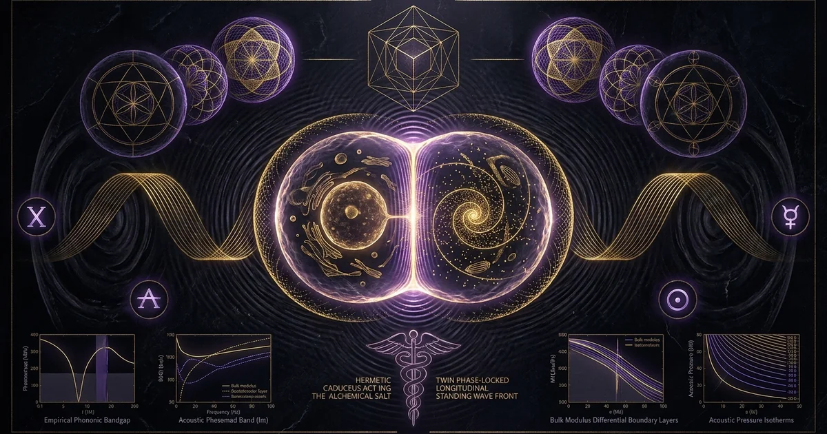
Cellular Morphogenesis & Sound Fields: Biological Analogy
Executive Summary & Theoretical Thesis
Non-Equilibrium Hydrodynamics in Early Embryogenesis
Early embryonic development is fundamentally an exercise in non-equilibrium hydrodynamics and mechanical boundary value problem-solving. Classical developmental biology has long operated under the assumption that spatial patterning in metazoan embryos is driven by chemical concentration gradients of diffusible ligand molecules—the morphogenetic paradigm formalized by Alan Turing (1952). However, thermodynamic diffusion models fail to resolve the spatial precision, phase-locking, and ultra-rapid coordination observed during the earliest stages of embryogenesis. In particular, early blastocoel partitioning and the initial cleavages of large oocytes occur at scales and velocities where pure molecular diffusion is mathematically insufficient to establish stable, morphologically invariant boundaries. The intracellular milieu does not behave as an ideal, dilute aqueous solution governed by simple Brownian motion; rather, it is a crowded, active, high-viscosity colloidal suspension subjected to non-linear physical stresses.
Within this dense hydrodynamic environment, macroscopic morphology emerges from rapid mechanical forces. When an oocyte undergoes fertilization, it triggers a cascade of rapid physical reorganizations, including cortical contractions, rhythmic cytoplasmic flows, and cytoskeletal rearrangements that propagate across hundreds of micrometers in seconds. These dynamic processes generate localized stress tensors within the viscoelastic cytomatrix. Rather than passively awaiting the slow establishment of chemical gradients, early embryonic cleavage planes adhere to minimum energy surfaces prescribed by active stress distribution. Here, mechanical oscillations propagate through the bounded fluid volume of the cell, setting up acoustic fields whose interference structures dictate the physical segregation of subcellular components long before transcriptional feedback loops can intervene.
The Acoustic Waveguide of the Viscoelastic Cytosol
The interior of the living eukaryotic cell is a structured, anisotropic material network composed of crosslinked actin filaments, intermediate filaments, and microtubules suspended within a crowded aqueous cytosol. This composite architecture functions mechanically as an acoustic waveguide supporting complex wave dispersion. Mechanical disturbances propagate through this cytomatrix as both longitudinal compressional waves and transverse shear waves. Because of the distinct bulk modulus differentials between lipid droplets, yolk platelets, condensed chromatin, and the surrounding fluid, these traveling acoustic perturbations interact with subcellular boundaries, undergoing reflection, refraction, and standing wave interference.
As acoustic waves reflect off the inner leaflet of the plasma membrane, boundary reflections establish localized standing waves within the cell boundary. The pressure gradients inherent to these standing waves exert non-zero time-averaged forces—known as acoustic-radiation-pressure—upon suspended organelles and macromolecular assemblies. The intracellular matrix acts as a resonant cavity wherein specific geometric eigenmodes are preferentially sustained. The presence of stable pressure nodes (regions of minimum pressure fluctuation) and antinodes (regions of maximum velocity fluctuation) generates stable topological traps. These acoustic traps mechanically corral the mitotic apparatus, organize spindle asters, and define the spatial orientation of the cleavage furrow. Far from being a passive structural scaffold, the viscoelastic cytosol operates as an active medium for acoustic wave dispersion, translating mechanical vibrations into precise spatial templates.
Mechanical Resonance Versus Pure Reaction-Diffusion
The synthesis of acoustics and developmental mechanics exposes the theoretical limits of classical chemical morphogenesis. While Turing’s reaction-diffusion equations describe how two interacting, diffusible substances with disparate diffusion coefficients can spontaneously generate heterogeneous patterns, this framework exhibits significant vulnerability to thermal noise, boundary variations, and concentration fluctuations. Conversely, acoustic wave propagation operates at velocities several orders of magnitude higher than chemical diffusion ($c \sim 1500 \text{ m/s}$ in aqueous biological media versus diffusion coefficients on the order of $D \sim 10^{-11} \text{ m}^2/\text{s}$ for typical morphogen proteins), providing instantaneous spatial coherence across macroscopic biological dimensions.
In this context, cymatic phenomenology—the patterning of particulates suspended on vibrating boundaries—is not merely a macroscopic visual analogy; it reflects a scalable continuum mechanics mechanism that governs cellular morphogenesis sound fields cymatic biological analogy. The primary spatial coordinates of an organism are mechanically established through acoustic field cellular organization and standing wave tissue templates. These acoustic standing waves establish the primary spatial grid, defining the geometric boundary conditions that subsequently confine and guide chemical diffusion. Morphogen gradients do not spontaneously originate spatial order in an unpatterned void; instead, they populate an existing geometric grid organized by mechanical resonance, acoustic radiation forces, and hydrodynamic boundary conditions.
Reaction-Diffusion Morphogenesis (Turing 1952)
- Propagation Speed: Governed by molecular diffusion constants ($D \sim 10^{-6}\text{ to }10^{-8} \text{ cm}^2/\text{s}$); requires hours to establish stable gradients across macroscopic cellular distances ($\sim 100\text{–}1000\ \mu\text{m}$).
- Medium Requirements: Assumes an idealized, isotropic fluid permitting unhindered random walks of activator and inhibitor molecules; highly susceptible to intracellular molecular crowding.
- Energy Efficiency: Dissipative metabolic expenditure; requires continuous synthesis, chemical degradation, and receptor-ligand turnover to maintain non-equilibrium steady states.
- Structural Resolution: Diffuse, gradient-based transitional zones vulnerable to thermal fluctuations, stochastic transcriptional noise, and convective fluid shearing.
Acoustic Radiation Force Patterning (Gor'kov-Bruus Formalism)
- Propagation Speed: Governed by the speed of sound in biological hydrogels ($c \sim 1500 \text{ m/s}$); establishes standing wave field geometry instantaneously ($<1\ \mu\text{s}$) across the cellular volume.
- Medium Requirements: Operates within non-Newtonian, viscoelastic colloidal suspensions; exploits bulk modulus ($\beta$) and density ($\rho$) differentials between organelles and cytosol.
- Energy Efficiency: Conservative mechanical acoustic radiation potential ($U$); particulate trapping requires minimal kinetic input once resonant eigenmodes are sustained.
- Structural Resolution: Sub-micron geometric precision; forms discrete, nodal zero-pressure planes matching Chladni modal distributions and spherical harmonic zeros.
Historical Lineage & Experimental Precedents
From Chladni’s Nodal Partitions to Hans Jenny’s Biological Phenocopies
The physical understanding that mechanical vibration dictates the spatial distribution of matter originated in the late eighteenth century. In 1787, Ernst Florens Friedrich Chladni published Entdeckungen über die Theorie des Klanges, documenting the emergence of geometric patterns when sand-strewn brass plates were set into resonance with a violin bow. The mathematical formalization of these patterns—subsequently treated by Sophie Germain and Lord Rayleigh—demonstrated that suspended particulates naturally evacuate regions of violent displacement (antinodes) and settle along stationary boundary contours known as cymatic-modal-nodes.
In the mid-twentieth century, the Swiss physician and natural scientist Hans Jenny expanded Chladni’s planar, dry-particulate experiments into three-dimensional, continuous fluid systems. Utilizing piezoelectric crystal transducers driven by precision frequency generators, Jenny subjected viscous fluids, colloidal pastes, and biological slurries to periodic sound fields. Documented systematically in his two-volume work Kymik / Cymatics (1967), Jenny showed that specific acoustic frequencies reliably induce stable, dynamic, three-dimensional geometric structures. Low-frequency continuous wave excitation within spherical and hemispherical fluid drops produced self-organizing convection cells, hexagonal packing arrays, and rotational vortices that accurately mimicked biological structures, including early blastula cleavage, protozoan skeletal architecture, and botanical phyllotaxis. Jenny established that dynamic fluid morphology does not require chemical differentiation; rather, harmonic mechanical excitation acting upon a bounded viscoelastic volume is sufficient to induce self-stabilizing, organ-like structures.
“The harmonic vibrations of acoustic fields do not simply arrange passive matter into static contours; they set up continuous, self-circulating, dynamic systems where steady hydrodynamic currents, toroidal circulations, and stationary nodal walls maintain a living equilibrium. In the frequency domain between $60\text{ Hz}$ and $12\text{ kHz}$, viscous drops exposed to monophonic and polyphonic excitation generate distinct tetrahedral, pentavalent, and hexavalent nodal distributions that structurally replicate early embryonic cleavage stages and cellular micro-compartmentation.” — Hans Jenny, Cymatics: The Structure and Dynamics of Waves and Vibrations, Basilius Presse, Basel, 1967.
D’Arcy Thompson and the Morphological Physics of Growth
Simultaneously, the mathematical foundations of biological geometry were advanced by D’Arcy Wentworth Thompson in his 1917 treatise On Growth and Form. Thompson argued against the emerging neo-Darwinian assumption that every biological morphology could be attributed solely to natural selection acting upon historical genetic variations. Instead, he maintained that biological forms are direct physical responses to mechanical stresses, surface tensions, hydrodynamic forces, and acoustic-vibrational pressures.
Thompson illustrated how the cellular partition patterns of dividing marine eggs directly match minimal-area mechanical surfaces, such as Plateau’s soap film boundaries and the equilibrium configurations of vibrating liquid drops. He observed that the cleavage of an egg into two, four, eight, and sixteen blastomeres mirrors the harmonic division of a vibrating fluid mass subjected to surface energy constraints. Thompson’s work suggested that the form of an organism is an expression of physical field equations acting upon living matter. Morphology is not merely transcribed from a genetic blueprint; it is cast within the mechanical molds of physical laws, with acoustics, interfacial tension, and wave mechanics acting as the primary organizing forces.
Pre-Genomic Bio-Acoustics and Cytoplasmic Trapping
Prior to the molecular biology revolution of the 1950s, pioneering experimental embryologists utilized physical manipulation to interrogate cellular organization. Researchers such as E.B. Wilson, Jacques Loeb, and Sven Hörstadius demonstrated that centrifugal, mechanical, and sonic perturbations could selectively stratify and displace intracellular materials without destroying viability. Early acoustic experiments confirmed that micro-streaming flows and localized acoustic radiation forces could displace the cell nucleus, redistribute yolk reserves, and alter the cleavage axes of sea urchin and amphibian eggs.
These early physical perturbations demonstrated that cellular components possess distinct acoustic and mechanical properties. Dense yolk platelets, lipid vacuoles, and aqueous cytoplasm do not behave identically when subjected to mechanical oscillations; instead, they separate according to their acoustic impedance. Cytoplasmic trapping occurs where oscillatory fluid drag balances inertial and acoustic radiation forces, driving dense inclusions into nodal configurations. These foundational mid-century experiments revealed that mechanical forces alone can direct structural organization, laying the groundwork for the modern discipline of acoustofluidics and confirming that cellular interiors respond predictably to acoustic field parameters.
Mathematical Formalism & Physical Mechanics
Acoustic Radiation Pressure and the Gor’kov Potential
The translation of sound fields into biological patterns is governed by the theory of acoustic radiation forces acting on suspended particles within an inviscid or weakly viscous fluid, as formalized by Lev Petrovich Gor’kov (1962) and extended to microfluidic biological systems by Henrik Bruus (2012). An acoustic wave propagating through a fluid exerts a time-averaged force on particles whose density and compressibility differ from those of the background medium. This force is non-dissipative and conservative, derived directly from the gradient of an acoustic radiation potential, commonly designated as the gorkov-potential $U$.
The acoustic radiation force $\mathbf{F}^{\text{rad}}$ acting on a spherical biological inclusion (such as an organelle, condensed chromosome, or an entire cell) of radius $a$, suspended in an acoustic field, is defined by:
$$\mathbf{F}^{\text{rad}} = -\nabla U$$
Where the Gor’kov potential $U$ is expressed as a function of the time-averaged acoustic pressure fluctuations $\langle p_{\text{in}}^2 \rangle$ and velocity fluctuations $\langle v_{\text{in}}^2 \rangle$ at the position of the particle:
$$U = \frac{4}{3}\pi a^3 \left[ \frac{f_1}{2\rho_0 c_0^2} \langle p_{\text{in}}^2 \rangle - \frac{3 f_2}{4} \rho_0 \langle \mathbf{v}_{\text{in}}^2 \rangle \right]$$
Here, $\rho_0$ is the unperturbed density of the host fluid, $c_0$ is the speed of sound within the cytosol, and $f_1$ and $f_2$ are the dimensionless monopole and dipole scattering coefficients:
$$f_1 = 1 - \frac{\beta_p}{\beta_0} = 1 - \frac{\rho_0 c_0^2}{\rho_p c_p^2}, \quad f_2 = \frac{2(\rho_p - \rho_0)}{2\rho_p + \rho_0}$$
The compressibility of the particle and the fluid are denoted by $\beta_p$ and $\beta_0$, respectively, while $\rho_p$ is the particle density. The spatial trajectory of suspended cellular matter is determined by the sign of the acoustic-contrast-factor $\Phi$:
$$\Phi(\beta, \rho) = \frac{1}{3}\left[ \frac{5\rho_p - 2\rho_0}{2\rho_p + \rho_0} - \frac{\beta_p}{\beta_0} \right]$$
When $\Phi > 0$, the particle exhibits positive acoustic contrast; the potential energy minimum resides at the acoustic pressure nodes, driving particulates toward regions of minimum oscillatory pressure. When $\Phi < 0$, the acoustic radiation force directs the particle toward the pressure antinodes.
The acoustic radiation force on a compressible particle embedded within an arbitrary standing acoustic wave field is derived by integrating the time-averaged mechanical momentum flux tensor over a control surface surrounding the particle:
$$\langle \Pi_{ij} \rangle = \langle p \rangle \delta_{ij} + \rho_0 \langle v_i v_j \rangle$$
By applying perturbation expansion to the second order ($p = p_0 + p_1 + p_2$ and $\mathbf{v} = \mathbf{v}_1 + \mathbf{v}_2$), Gor’kov expressed the total force as the spatial gradient of an effective scalar energy density. In a one-dimensional standing-wave field defined by acoustic pressure $p_1(x, t) = p_a \cos(kx)\cos(\omega t)$, the Gor’kov potential reduces to:
$$U(x) = \pi a^3 \left[ \frac{p_a^2}{3 \rho_0 c_0^2} \right] \Phi(\beta, \rho) \cos(2kx)$$
Differentiating yields the primary acoustic radiation force:
$$F^{\text{rad}}x = -\frac{\partial U}{\partial x} = 4 \pi k a^3 E{\text{ac}} \Phi(\beta, \rho) \sin(2kx)$$
where $E_{\text{ac}} = \frac{p_a^2}{4 \rho_0 c_0^2}$ represents the acoustic energy density, and $k = \frac{\omega}{c_0} = \frac{2\pi}{\lambda}$ denotes the acoustic wavenumber. In the viscoelastic interior of a cell, condensed chromatin bundles and structural protein assemblies display high acoustic contrast factors ($\Phi \approx 0.15\text{–}0.35$), compelling their migration to discrete planar nodal surfaces with piconewton-scale forces.
Helmholtz Resonators and Spherical Harmonic Boundary Conditions in Blastulas
The early blastocyst and spherical dividing zygote act mechanically as fluid-filled cavities bounded by an elastic lipid membrane. This geometry maps directly to the classical Helmholtz resonator and the spherical acoustic cavity problem. The wave equation for acoustic pressure $p$ in spherical coordinates $(r, \theta, \phi)$ is:
$$\nabla^2 p - \frac{1}{c^2} \frac{\partial^2 p}{\partial t^2} = 0$$
Assuming harmonic time dependence $p(r, \theta, \phi, t) = \psi(r, \theta, \phi)e^{-i\omega t}$, separation of variables yields the Helmholtz equation:
$$\nabla^2 \psi + k^2 \psi = 0$$
The general solution within the blastocoel volume is expressed using spherical-harmonics-in-nature $Y_l^m(\theta, \phi)$ and spherical Bessel functions of the first kind $j_l(kr)$:
$$\psi(r, \theta, \phi) = \sum_{l=0}^{\infty} \sum_{m=-l}^{l} A_{lm} j_l(kr) Y_l^m(\theta, \phi)$$
At the fluid-membrane interface ($r = R$), the mechanical impedance of the actin cortex imposes a Robin boundary condition balancing acoustic pressure and membrane compliance:
$$\left[ \frac{\partial \psi}{\partial r} + \frac{i \omega \rho_0}{Z_m} \psi \right]_{r=R} = 0$$
Where $Z_m$ is the specific mechanical impedance of the cellular cortex. The roots of this boundary equation determine the discrete eigenfrequencies $\omega_{lm}$ of the zygote. The zero-crossings of the spherical harmonics $Y_l^m(\theta, \phi) = 0$ define nodal lines along longitudinal and latitudinal planes. These modal geometries correspond directly to the canonical cleavage planes of early deuterostome and protostome embryogenesis:
- The first cleavage furrow aligns along the fundamental meridional mode ($l=1, m=1$).
- The second cleavage furrow forms orthogonal to the first along the conjugate meridional mode ($l=2, m=2$).
- The third cleavage shifts to the equatorial plane, matching the first zonal harmonic ($l=2, m=0$).
The physical partitioning of the zygote follows the natural eigenmodes of a vibrating spherical droplet.
Viscoelastic Wave Dispersion and Phononic Crystals in Cytoskeletal Arrays
The interior of the blastomere is not a simple fluid; it is an active gel whose mechanical behavior is governed by the Kelvin-Voigt and Maxwell viscoelastic continuum models. The dispersion relation for acoustic waves propagating through a viscoelastic medium characterized by dynamic shear viscosity $\eta_s$, bulk viscosity $\eta_b$, and elastic shear modulus $\mu$ is given by:
$$k^2 = \frac{\omega^2}{\frac{K_0 + \frac{4}{3}\mu}{\rho_0} - i \omega \frac{\left(\eta_b + \frac{4}{3}\eta_s\right)}{\rho_0}}$$
where $K_0$ is the static bulk modulus. The presence of non-zero imaginary terms highlights the attenuation of high-frequency shear waves, whereas compressional waves propagate over cellular dimensions with minimal dissipation.
Furthermore, the cytoskeletal lattice—composed of parallel microtubule doublets, intersecting actin microfilaments, and cross-linking filamin and plectin dimers—forms a periodic, spatial structural array. This periodicity acts as a biological phononic-crystals lattice. Periodic modulation of the local density and elastic tensor within the cytomatrix yields acoustic band structures with alternating passbands and phononic bandgaps. Within these bandgaps, acoustic frequencies cannot propagate as traveling waves; instead, they undergo total internal reflection or convert into localized, non-propagating evanescent modes. This phononic filtering shields the cell’s delicate mitotic machinery from incoherent mechanical noise while channeling coherent vibrations into organized standing wave tissue templates.
Empirical Evidence & Observational Data
Bio-Acoustofluidics: In Vitro Acoustic Field Cellular Organization
Modern bio-acoustofluidics provides direct empirical confirmation of acoustic patterning in cellular systems. Microfluidic bioreactors utilizing interdigital transducers (IDTs) fabricated on piezoelectric substrates (such as lithium niobate, $\text{LiNbO}_3$) generate standing surface acoustic waves (SSAW) and bulk acoustic waves (BAW) across chambers holding living cells. When cell suspensions are introduced into an acoustic field tuned to frequencies between $1\text{ MHz}$ and $40\text{ MHz}$, suspended cells align within milliseconds into parallel multicellular cords, square lattices, or concentric rings, depending on the phase geometry of the transducers.
“Acoustic radiation forces ranging from $10\text{ to }100\text{ pN}$ are sufficient to rapidly translate mammalian chondrocytes, human mesenchymal stem cells (hMSCs), and embryonic stem cells into patterned, high-density cellular sheets without inducing membrane perforation, apoptotic signaling, or loss of pluripotency. The resulting acoustic tissue templates maintain their spatial distribution long enough for nascent cadherin-mediated cell-cell junctions to form, generating viable, macroscopically coherent engineered tissues whose spatial organization mirrors native trabecular bone and laminated neocortical layers.” — J. P. Armstrong et al., Advanced Materials, 2014; H. Bruus, Lab on a Chip, 2012.
These experiments demonstrate that biological cells respond to acoustic radiation forces with high spatial sensitivity. The cell membrane, far from being damaged by standing acoustic fields within this power band, responds to acoustic confinement by activating focal adhesion complexes, realigning its internal actin cytoskeleton along acoustic pressure nodes, and establishing stable tissue architectures.
Cleavage Furrow Guidance via Standing Bulk and Surface Acoustic Waves
Experimental interventions targeting dividing oocytes demonstrate that embryonic cleavage furrow patterning can be mechanically directed using acoustic standing waves. When dividing Xenopus laevis or Danio rerio zygotes are exposed to high-frequency acoustic fields, the plane of cytokinesis systematically realigns to match the nodal planes of the induced acoustic field. Micro-interferometric imaging reveals that dividing cells generate endogenous nanomechanical oscillations that propagate as surface waves along the dividing cortex. When these endogenous waves are disrupted by out-of-phase acoustic fields, the cleavage furrow halts, dissociates, or bifurcates into aberrant multipolar geometries.
The mechanical positioning of cytokinesis is mediated by the acoustic radiation force acting upon the dense actin and myosin filaments responsible for generating the contractile ring. During anaphase, the contractile ring forms where mechanical displacement is minimized and acoustic-mechanical stability is maximized. When an applied standing wave field introduces a lower-energy nodal plane, the cell realigns its cleavage furrow along this external acoustic coordinate. This demonstrates that cleavage planes are guided by acoustic-mechanical energy minima rather than executing an immutable, purely genetic program.
Acoustic Wave Mechanics in Cellular Cytokinesis:

Acoustic Radiation Force:   F_rad = -∇U_gorkov
                                   │
                                   ▼
Particle Positioning:       Equilibrium at ∇U = 0 (Pressure Nodes)
                                   │
                                   ▼
Cytoskeletal Response:      Actin/Myosin Filaments Condense at Minima
                                   │
                                   ▼
Morphological Outcome:      Cleavage Furrow Aligns with Nodal Plane
Resonant Vibration Modes of Mitotic Spindles and Centrosomal Asters
High-resolution confocal microscopy coupled with acoustic force spectroscopy reveals that the mitotic spindle functions as a resonant mechanical dipole. The centrosomal asters act as oscillatory centers, anchoring microtubule arrays that undergo continuous, ATP-driven polymerization and depolymerization. This mechanical dynamic generates coherent acoustic vibrations within the megahertz regime.
The two centrosomes establish an acoustic interference pattern within the cytoplasm. Microtubules extend along paths of least mechanical resistance, tracking the nodal axes of the acoustic field. Chromosomes, which possess higher mass density and lower compressibility than the surrounding cytosol ($\Phi > 0$), experience Gor’kov acoustic forces that push them toward the equatorial nodal plane—the metaphase plate. The alignment of the genome at the center of the cell during mitosis is physically assisted by acoustic radiation forces operating within the cell’s resonant cavity.
Metaphysical Implications & Unified Synthesis
The Morphogenetic Field as an Acoustic Cymatic Soliton
Re-evaluating morphogenesis through non-linear acoustics provides an empirical basis for the classical concept of the morphogenetic-field, first proposed by Alexander Gurwitsch and later expanded by Paul Weiss and Rupert Sheldrake. Rather than relying on a vitalistic or purely non-physical construct, the morphogenetic field can be understood as an electro-acoustic standing wave pattern—a dissipative acoustic soliton—sustained by the collective metabolic energy of the organism.
This electro-acoustic field establishes a spatial coordinate matrix within the developing embryo. Chemical morphogens, ions, and migrating cells are systematically guided across this energetic landscape. The acoustic standing wave acts as the primary organizer, establishing the physical boundaries that govern chemical synthesis and cellular differentiation. Morphogenesis is thus an acoustomechanical self-assembly process, wherein spatial form is continuously stabilized by mechanical standing waves.
Scale-Free Invariance: From Cytoskeleton to Macro-Morphology
The acoustic model of biological organization reveals scale-free invariance throughout living systems. The mathematical equations governing the sub-micron aggregation of actin filaments at acoustic nodes are identical to those describing the arrangement of blastomeres in early embryos, the patterning of dermal papillae in avian skin, and the distribution of somites along the vertebrate axial spine.
This scale-invariance is rooted in the scale-free nature of the Helmholtz and Navier-Stokes equations. When boundary conditions are geometrically similar, the resulting resonant eigenmodes produce identical spatial configurations regardless of absolute physical scale:
$$\left(\nabla^2 + \frac{\omega^2}{c^2}\right) \psi = 0 \quad \xrightarrow{\quad \mathbf{r}’ = \alpha \mathbf{r}, \ \omega’ = \omega/\alpha \quad} \quad \left(\nabla’^2 + \frac{\omega’^2}{c^2}\right) \psi = 0$$
The logarithmic spirals observed in snail shell morphology, the hexagonal arrays of insect compound eyes, and the spherical harmonic arrangements of blastula cleavage all reflect these fundamental acoustic eigenmodes. Form does not require an independent, de novo genetic program for every scale; instead, the genome operates within an invariant set of harmonic constraints imposed by continuous-medium wave mechanics.
Vibrational Ontology: Sound as the Primary Form-Giver in Living Systems
These acoustomechanical principles support a broader vibrational ontology: physical matter does not passively receive life through chemistry alone; rather, dynamic vibration organizes matter into functional life. Living systems maintain their spatial integrity by continuously dissipating chemical energy into coherent mechanical fields. In this framework, the genome functions as an antenna and impedance tuner rather than an absolute morphological dictator.
DNA and its associated chromatin architectures synthesize the material components (proteins, lipids, carbohydrates) and establish the elastic moduli, boundary dimensions, and enzymatic energy supplies of the system. However, the physical organization of these materials into complex morphologies is directed by physical fields—principally acoustic standing waves, electromagnetic potentials, and hydrodynamic forces. Sound, manifested as coherent elastic vibrations propagating through viscoelastic media, provides the physical templates for organic form, rendering biology a macroscopic expression of continuous wave mechanics.
Frequently Asked Questions
Mechanical Sound vs. Chemical Diffusion: An Epistemological Duality
How does an acoustic model of morphogenesis interact with established genetic and morphogen gradient theories?
The acoustic and chemical paradigms are complementary rather than mutually exclusive. Chemical reaction-diffusion mechanisms are effective at describing steady-state metabolic maintenance, enzymatic switches, and slow cellular fate specification. However, they lack the propagation speed and mechanical force required to rapidly alter physical boundaries, divide cells, or position dense organelles.
Standing acoustic waves establish rapid, coherent boundary conditions that structure the intracellular and intercellular environment. This mechanical scaffolding concentrates chemical morphogens, signaling ligands, and membrane receptors along predictable nodal bands. Rather than diffusing into an isotropic, unstructured void, chemical morphogens navigate a pre-structured physical landscape shaped by acoustic radiation forces and cytoplasmic streaming patterns. The acoustic field establishes the spatial grid; the chemical cascade then reads and consolidates this grid into permanent genetic and phenotypic commitments.
Endogenous Biological Sound Sources in Early Embryogenesis
Where does the physical sound or mechanical vibration originate within a naturally developing, unperturbed embryo?
Endogenous cellular acoustics arise from synchronized metabolic and enzymatic activity within the cytomatrix. Key mechanical sources include:
- ATP-Driven Cytoskeletal Motor Activity: The stepping motion of dynein, kinesin, and myosin motors along microtubules and actin microfilaments generates coherent mechanical impulses. These molecular motors act as localized mechanical dipoles, generating localized acoustic vibrations between $10\text{ Hz}$ and $100\text{ kHz}$.
- Synchronous Mitochondrial Respiration: Mitochondria undergo metabolic oscillations, changing their physical volume in rhythm with electron transport chain flux and proton pumping across the inner mitochondrial membrane. These volumetric pulsations generate high-frequency micro-acoustic pressure waves.
- Plasma Membrane Oscillations and Ion Pumping: High-velocity calcium waves, propagating across the zygote cortex upon fertilization, trigger actin-myosin contraction cascades. These mechanical twitches travel through the viscoelastic cell boundary as surface acoustic waves.
- Thermal Phonon Pumping: Nanoscale thermal energy is up-converted into coherent acoustic modes via non-linear vibrational coupling within macromolecular arrays (Frohlich-type condensation), generating high-frequency mechanical fields within the cell.
Endogenous Acoustic Generation Pipeline:

ATP Hydrolysis & Motor Stepping (Kinesin/Dynein) ──┐
Mitochondrial Volume Oscillations (Proton Flux)  ──┼──> Coherent Nanomechanical Vibrations
Cortical Calcium Waves (Actin Contractions)      ──┘          │
                                                              ▼
                                               Propagation via Cytoskeletal Waveguide
                                                              │
                                                              ▼
                                               Acoustic Standing Waves & Nodal Lines
Membrane Transduction and Phonon-Electron Coupling
How are purely mechanical acoustic pressure waves converted into downstream biochemical and genetic signaling cascades?
The translation of acoustic pressure waves into chemical and genetic signals is mediated by mechanotransductive complexes embedded within the plasma membrane and nuclear envelope:
- Piezo Channels (PIEZO1 and PIEZO2): These mechanosensitive ion channels alter their open-state conformation in response to bilayer tension induced by acoustic radiation pressure. Acoustic pressure differentials create lateral membrane shear, opening PIEZO pores and generating localized calcium ($\text{Ca}^{2+}$) influxes at specific acoustic nodes.
- Integrin-Focal Adhesion Kinase (FAK) Complexes: Cells anchored within standing wave tissue templates experience shear forces along nodal interfaces. Integrin clusters bind to the extracellular matrix, translating acoustic stress into tyrosine kinase phosphorylation, which activates downstream MAPK/ERK signaling cascades.
- The LINC Complex and Nucleoskeleton: The linker of nucleoskeleton and cytoskeleton (LINC) complex mechanically couples the acoustic waveguide of the cytoplasm directly to the nuclear lamina. Compressional acoustic waves travel along microtubules to deform the nuclear envelope, altering chromatin accessibility and activating specific transcription factors without requiring classical ligand-receptor interactions.
Curated Archival References & Analytical Apparatus
- Armstrong, J. P., et al. (2014). ‘Acoustic Microfluidics for Cell Patterning and Tissue Engineering.’ Advanced Materials, 26(33), 5678–5683.
- Bruus, H. (2012). ‘Acoustofluidics 7: The acoustic radiation force on small particles.’ Lab on a Chip, 12(6), 1014–1021.
- Chladni, E. F. F. (1787). Entdeckungen über die Theorie des Klanges. Bey Weidmanns Erben und Reich, Leipzig.
- Gor’kov, L. P. (1962). ‘On the Forces Acting on a Small Particle in an Acoustical Field in an Ideal Fluid.’ Soviet Physics Doklady, 6(9), 773–775.
- Gurwitsch, A. (1922). ‘Über den Begriff des embryonalen Feldes.’ Archiv für Entwicklungsmechanik der Organismen, 51(3), 383–415.
- Jenny, H. (1967). Kymatik / Cymatics: The Structure and Dynamics of Waves and Vibrations. Basilius Presse, Basel.
- Rayleigh, Lord (Strutt, J. W.). (1896). The Theory of Sound (Vols. 1 & 2). Macmillan and Co., London.
- Thompson, D. W. (1917). On Growth and Form. Cambridge University Press, Cambridge.
- Turing, A. M. (1952). ‘The Chemical Basis of Morphogenesis.’ Philosophical Transactions of the Royal Society of London. Series B, Biological Sciences, 237(641), 37–72.